Welcome to Anti Aging Fruits

Opening Hours : Monday to Saturday - 8am to 9pm
  Contact : (760) 481-8800

All Posts Tagged: HGH For A Longer Life

Risks Associated with Low HGH in Men

Men are extremely different than woman when it comes to the risks which arise from low

(HGH) Human Growth Hormone. Men with risky low levels of HGH will suffer from

extraordinary lack of energy and motivation.

HGH in men is exceptionally crucial as the man’s two most significant hormones which build

them up physically and even emotionally are HGH and Testosterone. Women are much more

complicated hormonally speaking.

Please let us make this very clear Protein has been scientifically proven to build human beings

up. Therefore we call the building up process Anabolic…which has NOTHING to do with

steroids…HGH is NOT a steroid but it is the single most important hormone in a man’s body.

Conversely, there are substances that tear the body down and that is called the Catabolic

process and this is highly dangerous for men to be in a catabolic state, ever.

Simply put, if a man is low in HGH he is automatically catabolic.

What exactly does that mean and just how dangerous a risk is this?

It means that when a man is in a catabolic state from low HGH he is not able to replace cells

and tissues faster than his body is breaking them down. What exactly does that mean?

According to Dr. Sam Baxus, one of the world’s foremost authorities for over 40 years on low

HGH…it means that you as a man are needlessly slowly dying faster than their male friends who

do not yet have low human growth hormone blood levels.

There also exits a direct relationship between HGH and testosterone in men such that if a man

is low in HGH it is very likely to have a negative impact Testosterone production. Thus more

muscle weakness, fatigue, more fat lower lean muscle mass, and depression in men.

It means, that every aspect, every part of your body is being torn down piece by piece.

And the leading first symptom for men is lack of energy and low motivation.

Then men notice an increase in fat around the belly area and a loss of tone to their muscles.

This alone is a risk which is certainly not acceptable once a man understands that inner fat

increased around the waistline has clearly been established by cardiologists as an INCREASED

risk of having a sudden and severe Heart Attack.

Demographic studies at the University of North Carolina (chapel Hill) often referred to as the

little Harvard of the South… is known to produce the best studies of risk for illnesses of every

type according to age and the numbers of folks in each age group.

Regarding their study on HGH by age groups something enormously profound was gleaned.

Age 18, is the best age group nationwide according to HGH levels and the almost complete lack

of illness in that age group. We noticed that every 8 years after age 18 there was and is a

dramatic rise in age related disease so if one takes age 18 as nearly perfect for HGH purposes

and we times 18 by 3 we get age 54 the highest group of men by age for heart attacks each

year. Between age 45 and 55 men are at severe risk for heart attacks

Thus if we look at all age related disease due to lack of HGH by this best demographic study

ever done so far, we find an enormous mistake. Generally it has been said we lose 50% of our

HGH output every 30 years past age 20. So it has been said for years that age 50 you make only

half the HGH as you did at 20

BUT if one delves deeply into the University of North Carolina’s massive demographic research

study on age related illness and relationship to HGH levels we easily see that after age 18 we

lose 50% of our HGH output every 8 years after the age 18.

So for men the risks come on generally around age 35 to 40 with belly fat and lack of energy

and motivation.

Next there are changes in the make-up of cholesterol with an increase in the bad LDL and also

an increase in the dangerous Triglyceride levels going up for men.

A man’s stamina decreases when HGH levels are low.

Men experience less lean muscle with low HGH

Men also complain about being more sensitive to heat and cold with low HGH. Men are more

sensitive to light. Especially bright light, which is an indication of breakdown of eye tissue.

Men experience as an early warning sign of low HGH a lack of sexual desire and function.

Eventually, bones density becomes more porous and fractures from a simple trip fall can occur

The immune system starts to breakdown for men if they do not take action and get themselves

on more optimal youthful blood levels of HGH.

Men, you can forgo all these dangerous risks. AAI provides true expert advice and expert HGH

treatment protocols. Women live 10 years longer than men. Come on guys 10 years is a mini

lifetime.

Read More

Is it time for a new paradigm in mental health?

Are non-drugs, like virgin coconut oil, a safer and more effective alternative to stress and depression?

A new study conducted in Malaysia looked at the effects of consuming high-antioxidant

virgin coconut oil on mental health.

Published in the journal Experimental and Therapeutic Medicine and believed to be the

first study of its kind, researchers evaluated the anti-stress and antioxidant effects of

virgin coconut oil in mice with stress-induced injuries. The title of the study is “Anti-

stress and antioxidant effects of virgin coconut oil in vivo.”

The researchers performed several stress tests on groups of mice. Control groups

included untreated mice and mice not subjected to stress and virgin coconut oil was

compared to a commonly prescribed psychiatric drug, Diazepam.

Their results were quite impressive, and suggest that using a high quality virgin coconut

oil can rival antidepressant drugs without the dangerous side effects. The researchers

attributed the success in treatment to the unique mixture of medium chain fatty acids

found in coconut oil rich in saturated fats, and to the antioxidants present in higher

grade, less processed virgin coconut oils.

While we do not endorse the supposed “science” behind psychiatric drugs, which

attempts to measure such things as “neurotransmitters” and “biochemical profiles” as

true indicators of mental health that can be altered by chemical drugs, it is encouraging

to see researchers consider natural foods as alternatives, given the fact that they do

not have all the serious side effects that psychiatric drugs do.

One of the more interesting tests conducted in this study was a measurement of

“immobility time” after a forced swim test. The researchers found that the untreated

mice had a longer immobility time than mice treated with virgin coconut oil. They

attributed this to the high medium-chain fatty acid content of coconut oil, which is

known to produce thermogenesis and increased energy.

One area where virgin coconut oil (VCO) really outperformed the drug Diazepam was in

the area of oxidation and elimination of free radicals. This is something that can be

measured with lipid peroxidation (MDA) and antioxidant enzyme SOD levels. Stress is

known to increase oxidation and the creation of free radicals, leading to neuronal cell

damage and death. Antioxidants, on the other hand, reverse this trend and help

prevent further neuronal damage.

The researchers found:

VCO was able to reduce lipid peroxidation and increase the activity of SOD in the serum

of mice undergoing the forced swim test and the brains of mice subjected to chronic

cold restraint. It was previously reported that VCO is rich in polyphenols and these

antioxidants may contribute to the increased levels of antioxidant enzymes, which

subsequently reduce lipid peroxidation and inflammation in VCO-treated mice.

Restoration of antioxidant levels in the brain may help prevent further neuronal damage

and avoid subsequent depletion of monoamines, including DA. In conclusion, the

present study demonstrated the potential of VCO in preventing exercise- and chronic

cold restraint stress-induced damage and restoring the antioxidant balance. This

promising anti-stress activity may be attributed to the polyphenols and medium-chain

fatty acids present in VCO.

It is high time for a new paradigm in mental health. Drugs are not the solution to stress

and depression. Non-drug alternatives are not only safer, but can be more effective

than pharmaceutical drugs as well.

Reference

Anti-stress and antioxidant effects of virgin coconut oil in vivo.  Experimental and

Therapeutic Medicine – Jan 2015; 9(1): 39–42

Read More

Reversing Atherosclerosis and Cardiovascular Disease

Healthy senior couple over fresh fruits and vegetables background.

Healthy senior couple over fresh fruits and vegetables background.

by: Larry Sosna N.D. PhD HHP

The Medical establishment would have all of us believe that coronary heart disease is irreversible and must lead to

either the eventual so called heart attack due to clogged arteries; or to by-pass operations, stents, followed by

balloon angioplasty. They tell us with Statin drugs the patient will then be ok. The statistics paint a very different

picture. Two out of every three people who have had cardiac by-pass surgery will need it again within 10 years.

Ten out of ten cardiovascular patients who have had stent procedures will need them again within 6.4 years.

Clearly, even with massive doses of statin drugs the treatment protocol for cardiovascular patients in use today by

the medical establishment is a failure in the sense that it’s not a cure and the above protocol is an ongoing radical

process for the body to endure.

One need  look no further than the case of former President Bill Clinton who had Quadruple by-pass heart surgery,

stent after stent, balloon angioplasty again and again, massive statins and still he had advanced coronary artery

disease(CAD) until he finally discovered a natural, yet very scientific integrative treatment protocol which has

reversed his CAD.

In the 1960’s and 1970’s Nathan Pritikin M.D. developed a unique diet rich in massive amounts of fruits and

vegetables and very low in total calories and fats. Dr. Pritikin developed this protocol after he was diagnosed with

severe atherosclerosis of the coronary arteries. He was so convinced his protocol reversed CAD he left instructions

in his will that when he died he was to have a cardiologist examine the condition of his arteries. Meanwhile

thousands of his patients went off all medications and never had any more coronary artery problems. After Dr.

Pritikin died in 1987, his heart was dissected; the arteries were determined by cardiologists to be clean and in the

condition of a 25 year old.

In 1987, two years after Dr. Pritikin’s death, the Journal of the American Medical Association announced a study

that showed the reversal of atherosclerosis in the coronary arteries of humans who were on the Pritikin protocol as

administered at the Pritikin Longevity centers had been a success.1 Numerous subsequent studies confirmed Dr.

Pritikin was scientifically correct and the medical establishment’s position fatally flawed.2,3

Tens of millions of Americans needlessly perished because the medical establishment refuses to understand the

true causes of Coronary Artery Disease.

The over-promotion of “Statin” drugs has resulted in today’s cardiologists focusing on getting their patients’ total

cholesterol and LDL down to dangerously low levels. Pharmaceutical company advertising has made it appear as if

the only cause of CAD is excess LDL and cholesterol; however over a thousand scientific peer reviewed research

studies show this conclusion to be completely false…

Beginning in 1979, medical researcher scientists made discoveries which clearly indicated that CAD was the result

of the oxidation of LDL that results in the formation of blocked arteries and arterial damage.4,5   Thousands of peer

reviewed studies now reveal how oxidized LDL creates CAD from start to finish.

There are doctors who argue that CAD is all about inflammation and they are correct but what they failed to

realize is that oxidized cholesterol mainly LDL injures endothelial cells and causes this massive inflammation of the

coronary arteries.6-12

Oxidized LDL causes endothelial cells to secrete ultra-sticky adhesion molecules that allow white blood cells to

penetrate the inner lining of the artery. This is where initial fatty streaks congeal and atherosclerotic plaques

develop in thickening layers.13

Oxidized LDL turns on white blood cell gene expression that enable them to convert into foam cells, which results

in continuous accumulation of oxidized LDL and thick plaque in the coronary arteries.14

Oxidized LDL initiates an inflammatory process by causing foam cells to make molecules that attract

proinflammatory cells and cytotoxins that further inflame the artery linings.15

Oxidized LDL enhances the process whereby immune cells, foam cells, smooth muscle cells, and the lining of

coronary arteries called endothelial cells, degrade collagen, which leads to the rupture of the fibrous plaque. The

extreme heat from the inflammation can cause even small amounts of cholesterol to blow up 40 times their normal

size and clot up an entire coronary artery, thus recently the Harvard study group called this phenomenon Popcorn

Cholesterol. This finally explains why 53% of all fatal heart attacks occur in people with rather low cholesterol,

something until recently which has greatly confused cardiac medical researchers.16

Forty years ago coronary artery disease was the #1 cause of death in America roughly 600,000 people died each

year from CAD. Today despite doctors handing out statin drugs like candy CAD is still the #1 cause of death each

year at roughly 630,000 deaths per year. We have the highest rate of cardiac disease in the entire world despite all

of the most so called state of the art drugs…in fact statin drugs have led some 200,000 people to develop

irreversible muscle wasting syndrome which is always fatal; and statins have also been linked to advanced liver

and kidney disease.

THE SOLUTION

In reality the solution to this problem of oxidized LDL is very simple if you understand plant based biochemistry.

Dr. Pritikin thought the reversal of plaque and CAD was due to the very low intake of fats in the diet he created. In

fact that had little to do with anything, it was the antioxidants from a huge assortment of fruits and vegetables

which prevented LDL from oxidizing in the coronary arteries and thus stopped inflammation and the whole above

described process of oxidized LDL leading to CAD.

The problem with the Pritikin program is it is so very restrictive in calories that most folks simply cannot make a

lifelong commitment to it. We also now know about antioxidants in the form of nutraceutical plant based

supplements 1000’s of times more powerful then eating the fruits and veggies on the Pritikin diet.

We should all breathe easier knowing that when it comes to INHIBITING LDL oxidation we can now stop it cold with

natural biochemicals such as UBIQUINOL CoQ10…. With the exception of Alpha Lipoic Acid and the active

biochemical in Pomegranate (Proanthocyandids, and Polyphenols) nothing is effective as CoQ10, we can actually

now improve the inhibition of oxidized LDL cholesterol by 130%… far greater protection then the Pritikin diet.17

Yet we can go even far greater in reversing CAD by taking Nutraceutical quality Omega 3 oils every day. Omega 3

fish oil, Krill oil and Green Lipid muscle oil are extraordinarily powerful inhibitors of inflammation. The above are

such powerful High Density Lipids the good HDL’s, they can virtually assure that hard LDL cannot clump together,

in fact Eskimos and the arctic circle indigenous peoples account for five million people. They eat huge amounts of

cholesterol from seal blubber and whale blubber yet we have never found even one upon autopsy who had CAD.

Think about that, although they eat a diet 80% pure cholesterol they are completely protected by the 18% of their

diet which is cold water fish oil in the form of wild salmon.18-22

Stacking Powerful Antioxidants… The Sosna Protocol to cure advanced CAD

I have created a unique program by stacking the most powerfully effective antioxidants to reverse CAD for even

the most advanced CAD patient. Besides the above mentioned powerful antioxidants the stacking of nutraceutical

antioxidants also includes:

SAMe, Folic acid, isoflavones, Epigallate-Catchen-3 Galate, Resveratrol, Tocotrienols, Vit. E, Lutein, Lycopenes,

Ascorbyl Palmitate, Gingerols, Silymarin, Bromelain, Calcium D-Glucarate, Punicalagins, Hesperidin, Canthaxathin

and several others if need be. All of these super antioxidants are found only in the bio-diversity of plant based

molecular biochemistry in nature and in scientifically fresh cold pressed ocean based omega 3 fatty acids.

President Clinton is receiving with stellar results a similar protocol to mine…He will not need any more balloon

angioplasty or any other invasive treatments again so long as he stays on this treatment protocol for life.

In addition we have discovered plant extracted sterols can lower LDL by 38% in a very unique manner. They trick

the production of harmful LDL from entering the blood stream and instead send them a signal to enter the large

intestines where they are excreted via normal bowel movements.

In conclusion we have had for a long time the most effective all natural treatment protocol for reversing

cardiovascular disease. All the natural biochemicals I have mentioned are derived from plants, fruits, and marine

life, extracted and highly concentrated by a leading biotechnology company which is ISO 9001 and ISO 17025

While one may be tempted with this information to do it yourself without expert guidance, I strongly urge you to

consider the following. Eighty percent of all stores bought cold water fish oil sold as omega 3 HDL is already rancid

when purchased! Rancid oils have enormous amounts of free radical oxidants which cause CAD.  Unless you are

positive you are obtaining your nutraceutical supplements from a true ISO certified biotechnology laboratory be

very careful. The quality and purity of store the supplements will be no more than 20% pure due to their poor

quality extraction methodology, the other 80% may include large amounts of biological fragments, fillers, and

environmental toxins to the point where one is actually consuming enormous amounts of free radical oxidants

which seriously damage the heart, and other organs! Why? Because market competition is so fierce in the

supplement industry over 90% of the drug store and health store brands buy their raw materials from China; where

raw materials for making supplements are extremely inexpensive. The problem is China is the most polluted

industrial toxic country and raw organic material from China to make supplements will have high levels of toxins.

Any supplements made in the USA but sourced from China may be highly toxic. They will never be made by ISO

9001 ISO 17025 biotechnology companies; you simply cannot find this highest quality in health food stores and if

they claim they carry such a high standard ask the manager for a certified copy of ISO 9001 ISO 17025 for the item

you desire to purchase. They cannot produce the above mentioned certifications because there are only several

biotechnology companies worldwide, that have both ISO certifications and they only sell to doctors, dentists,

naturopathic doctors and nutritionists with a PhD.

Peer Reviewed research and footnotes.

1. Roberts CK, Chen AK Bernard JR Effects of short term diet and exercise intervention in youth on atherosclerotic

disease risk factors. Atherosclerosis. 2007 mar; 191 (1) : 98-106

2. Roberts CK, won D, pruthi S, et al. Effects of short term diet and exercise intervention on oxidative stress,

inflammation, MM-9, and monocyte chemotactic activity in men with metabolic syndrome factors. J Appl

Physiol. 2006 may; 100(5): 1657-65.

3. Roberts CK, Vaziri ND, Bernard RJ. Effects of diet and exercise intervention on blood pressure, insulin,

oxidative stress, and nitric oxide availability. Circulation. 2002 Nov 12;106(20):2530-2.

4. Anderson JW, Konz EC, Jenkins DJ. Health advantages and disadvantages of weight-reducing diets: a computer

analysis and critical review. J Am Coll Nutr. 2000 oct;19(5):587-90

5. Henriksen T Mahoney EM Steinberg D. Enhanced macrophage degradation of low density lipoproteins previously

incubated with cultured endothelial cells: recognition by receptors for acetylated low density lipoproteins.

Proc Natl acad Sci USA. 1981 Oct;78(10):6499-503.

6. Hessler JR, morel DW, Lewis LJ, Chisolm GM. Lipoprotein oxidation and lipoprotein-induced cytotoxicity.

Arteriosclerosis. 1983 May;3(3):215-22.

7. Quinn MT, Parthasarathy S, Fong LG, Steinberg D. Oxidatively modified low density lipoproteins: a potential

role in recruitment and retention of monocyte/macrophages during athero-genesis. Proc Natl Acad Sci USA.

1987 may;84(9):2995-8.

8. Ross R. Atherosclerosis—an inflammatory disease. N Engl J med. 1999 Jan 14;340(2):115-26.

9. Ross R. The pathogenesis of atherosclerosis-an update. N Engl J Med. 1986 Feb 20;314(8):488-500.

10. Yla-herttuala S, Palinski W Rosenfeld ME, et al. Evidence for the presence of oxidatively modified LDL in

atherosclerotic lesions of rabbit and man. J Clin invest. 1989 Oct;84(4):1086-95.

11. Steinberg d, Carew TE, khoo JC, witztum JL. Beyond cholesterol. Modifications of LDL that increase its

atherogenicity. N Engl J Med. 1989 Apr 6,320(14):915-24.

12. Berliner JA, navab m, Fogelman AM, et al. Atherosclerosis: basic mechanisms. Oxidation. Inflammation, and

genetics. Circulation. 1995 May 1;91(9):2488-96.

13. Suits AG, Chait A Aviram M, Heinke JW. Phagocytosis of aggregated lipoprotein receptor-dependent foam cell

formation. Proc Natl acad sci USA. 1989 Apr;86(8):2713-7.

14. Zhang WZ, Vernardos K Finch S, kaye DM. detrimental effect of oxidized LDL on endothelial arginine

metabolism and transportation. Int J Biochem Cell Biol. 2008;40(5):920-8

15. Faloon W. A lethal misconception of epidemic proportion. Life Extension. 2007 May; 13(5):7-14.

Danesh j, Lewington S, Thompson SG, et al. Plasma fibrinogen level and the risk of major

Read More

Glycomics, One of the 10 emerging technologies that will change the world.

MIT said the field of Glycomics, is “One of the 10 emerging technologies that will change the

world.” Notice, they did not say, one of the 10 medical technologies but one out of ALL

technologies that will change our world. It is as important as DNA and the Genome because the

8 major fundamentals of Glycobiology control both DNA and RNA. We even have the

Glyconome! Glycobiology controls our hormones and how they function…If you cannot get

enough of these 8 essentials life sustaining Glyconutrient’s, no matter how much HGH you take

it will have no effect or a neg. effect I am one of the few true experts in this exploding field. The

value to this more ramped up scientific language is so unique in this article that Google will

get in on these words I am using, KEY in on them and our standing in Google will go up

and up because no other competitor knows the scientific language of this anti-aging

breakthrough.. but Google’s algorithm’s and scrubbers places a very high value when you

write what others cannot or have not. When it is singular, as mine is and only on our

website blog Google loves that! That is why I want to write several articles on this topic so

germane to regeneration and living heathier for much longer. AND, there is big money in

this field selling sugars that heal. Please let me know what you think.

HELP DEFEAT ILLNESS AND GREATLY EXTEND YOU’RE LIFESPAN

Seminal Research by Larry Sosna N.D. PhD HHP

GLYCOBIOLOGY OF LIFE

See and hear what the true great university scientists are saying. “This is the future

today”, declares Dr. Gerald Hart of John Hopkins University…The number one rated

medical/scientific teaching hospital in the entire USA. “We won’t understand

Immunology, Neurology, Developmental Biology or Pathology until we get a handle on

Glycobiology.” ….. “If you ask, what is the Glycome for a single cell type it will be many

thousands of times more complex than the genetic Genome,” says Ajit Varki past

director of the Glycobiology Research and Training Center at Cal Tech. Professor

Raymond Dwek, head of the University of Oxford’s Glycobiology Institute, who

coined the term Glycobiology in 1988 says, “ As recent advances in genetics have

unfolded, the importance of sugars which heal has become ever more apparent.”…..

Varki said, “It’s like we just discovered the continent of North America. Now we have to

send out scouting parties to find out how big it is…..”

New Scientist Magazine, October 2002

I’m certain you have been told many times, sugar is one of the main enemies in life

causing all kinds of disease states and folks that can indeed be true. There are harmful

sugars and there are sugars so vitally important to vigorous good health that without

them we would all have suffered and died a long time ago. In fact, without these

miraculous special sugars we never would have made it out of infancy.

Glyco means sweet, there are bad sugars and ones we cannot live without, they are the

healthy Glyconutrients. If you are on one of those fad diets that does not allow the life

enhancing complex carbohydrates then you cannot make Glycoproteins which are

molecules that combine sugars with proteins as well as glycolipids, which are

combinations of healing complex sugars with fats. The term for these life giving

combinations are called glycoconjugates. These glycoconjugates have brought together

the world’s most brilliant medical scientists to comprehend a field so sophisticated the

combinations of glycoconjugates are infinite in how they arrange themselves to yield a

healthy body free from disease if you have them in high enough blood levels.

It will be useful to provide one with a high level of education concerning the immense

healing powers of a strong personal Glycobiology. Let’s shine some light on what

healthy levels of Glycoconjugates can do for the human body to improve both structure

and function.

Immune System Modulation:

Glyconutrients are necessary for healthy immune cells and a body wide capable

immune system function. They have been shown to:

Play a key role in many aspects of cell and tissue regeneration and repair, as well as

cell survival. They have strong positive effects on asthma and allergies in general.

Glyconutrients are one of the very few methods of preventing and or slowing down even

rheumatoid arthritis, lupus, and improves the symptoms of periodontal disease, canker

sores and herpes simplex 1 of the lips.

They have a favorable outcome in suppressing skin reactions and contact dermatitis as

well as inhibiting bronchitis. Many studies show they can prevent arthritis, substantially

reducing pain and increasing joint mobility in osteoarthritis, the most common form of

arthritis.

CANCER RESEARCH:

Glyconutrients and glycoconjugates help to inhibit growth and or tumor cell metastasis

in certain types of cancer. They do this by instructing white blood cells called Natural

Killer Cells to mobilize and seek out and destroy cancer cells. Today there is an entire

field of Glycobiology devoted to cancer research and the NIH and CDC have received

hundreds of millions in government funds to further enhance the field of Glycobiology

and cancer control.

Stress

An adequate supply of dietary glyconutritional healing sugars is vitally important during

periods of body wide stress, since glycoconjugates synthesized from eight key sugars

play key roles in many aspects of tissue healing and repair as well as cell survival

during extremely stressful periods. Corticotropin-releasing factor receptor is hormone

glycoprotein that regulates responses to emotional and other types of stress by

coordinating the endocrine, behavioral and immune responses to stress through

hormonal actions in the brain.

The Heart and Glycobiology

Glycolipid Conjugates are responsible for correct Low Density Lipid receptor site

instruction…These Low Density Lipids are the kind of cholesterol that can cause a

blocked artery and thus a heart attack…. But regulation of the dreaded LDL’s by

Glycolipid Conjugation makes the likelihood of artery blockage by LDL’s a very unlikely

event.

Most of us have heard that cold water fish oil is protective to the arteries of the heart.

When we all have high levels of Glycolipids the Omega 3 fatty acids become like a

super biological Teflon…coating the linings of the arteries so the bad types of LDL

cholesterol cannot aggregate and build up on the artery linings from both sides of the

coronary arteries thus in the scientific review of many research cardiologists who have

researched Glycolipid conjugation they have published findings that are very highly

suggestive that proper amounts of Glycolipids in the blood protects against a buildup of

bad LDL aggregation, such that the arteries seem to be fully protected from a heart

attack. The problem is a mass of Americans do not have adequate blood levels of

Glycolipid Conjugates especially, Galactose which is of great importance for normal

heart and brain health.

Glyconutritional high levels of galactose, mannose, galactosamine and sialic acid binds

vitamin B12 to make B12 bioavailable to the cells. In other without the above mentioned

healing sugar combinations you can inject all the B12 in the world and it will not work.

The same combination of Glucoconjugates helps people from drug and alcohol cravings

all of which are injurious to the heart and liver.

AND the exact same above described combination, seriously reduces HUNGER

Hormone Function

Gonadotropin hormones are glycoproteins derived from the pituitary gland which

controls the release of many other hormones throughout the body including human

growth hormone (hgh).

Glycosylation of gonadotrophin hormones affects their size, circulatory life span, ease

of movement through cells, storage and secretion, clearance, immunoreactivity and

hormone bioactivity within the entire human body.

Glycosylation of IGF receptors contributes to their tissue regeneration and tissue

differences, which affects their biological activity. If an individual does not have

adequate blood levels of Glycoconjugates (the healing 8 combined sugars) all hormone

activity no matter how much one takes will be seriously insufficient to do their job in the

human body and that can be a health crisis.

Glycoconjugation which does not occur in its proper sequence will lead to a disruption

of insulin formation and correct synthesis and can be causal in some types of diabetes.

The sugars I am about to share with you are so incredible they actually have a

language all of their own. I call them the great hidden code of human life itself. They are

every bit as important as DNA. Amazingly, when these life giving sugars are combined

they can even change mistakes in DNA expression. Mistakes… that can lead to cancer

and every other nightmare type of nightmare illness. However, I am going to show you

how to take a true and certain quantum leap in your ability to regenerate, fight off

virtually all illness, protecting you’re cells, tissues and all of the organs, collectively in all

of our bodies.

You may be thinking this is absurd, sugar is bad for us, correct? Not so simple, just the

sugar D-Ribose is so vital to life that without it we cannot make Messenger RNA and

without messenger RNA you may as well not even make DNA because both are

needed for correct structure and proper functioning of each and every cell, tissue and

organ our bodies. You might even remember from high school biology in the 9th grade

that RNA stands for Ribo Nucleic Acid…and it’s very back bone comes from our good

friend, the amazing D-Ribose SUGAR.

I am very excited to share this science with you. Unfortunately this is a science within

the medical field that is so complex it was not until the 1960’s to 1980’s that we got our

arms around this field of Molecular Glyco-Synthesis. And just like the field of DNA it took

the most brilliant hand full of scientists in molecular bio-science to advance this new

field and show how massively important it is. The field is so important to every phase of

our life cycle; I still study it weekly even though it has been a 20 years endeavor. That is

how important this field is and it is an honor to share this life enhancing information with

you, our family of friends here at AAI …. Though the field to this day is not being brought

to the public with much attention or consistency.

We have given you a small taste of the importance of the 5 carbon sugar D-Ribose now

let’s really delve into the Art and Science of the other life giving sugars.

There are eight Essential Sugars called Saccharides required for Glycoprotein

Synthesis…They are as follows Xylose, Fucose, Mannose, Galactose, Glucose, N-

Acetylglucosamine, N-Acetylgalactosamine, and N-Acetylineuraminic Acid.

What does this mean? It means no expression of protein’s abilities to build up the cells

into tissues and then into organs can occur. No Protein, no big muscles no matter how

hard you work. Actually, no protein , means death That means that without these eight

amazing sugars which create Glycoprotein Synthesis we would not have a body to even

take care of. That is why you must learn how to obtain these super regenerative eight

sugars…if you want to be in a perfected state of excellent health.

Guess what else? We cannot make any HORMONES or even utilize them efficiently

without these super eight sugars but wait there is more good news. Since the early

1990’s Gluco-Scientists found 200 other super sugars from plant based sources,

funding to scientifically evaluate all 200 found so far has several years ago reached the

many hundreds of millions of dollars so are discovering the roles of all 200 sugars.

Mother Nature’s vast array of biochemistry, including these 200 sugars has a specific

purpose. The overwhelming majority of them will be to create an even more complex

language to further healing, cell regeneration and repair. So please don’t be stuck on

eating (sucrose white ultra-refined table sugar) as sucrose in that form is BAD for

human consumption.

Speaking of Hormones, about 10 years ago I learned from Dr. David Wesser M.D.

DDS, a very close friend and teacher, taught if you combine a significant dose of D-

Ribose with Galactose, the human body will regenerate all hormones that have reached

the end of their life cycle, bringing them right back to the top of their life cycle. If you

want to feel like a Greek God, put this into you’re program.

Remember the eight super sugars are a code containing an extensive language. Each

sugar is like a 1000 page book in terms of its ability to communicate instructions

throughout the entire body. Each page in any of the eight books can code and give

hundreds of millions of directions throughout the body. So, we are talking about a code

of life capable of issuing Trillions of life enhancing instructions from the molecular level

to the cells, tissues and organs and even the brain itself gets its instructions to develop

into a brain, as well as sustain you’re brain.

For Illustrative purposes, these molecular communication codes are like precisely

shaped words that protrude from the cells surface and are recognized and understood

by neighboring cells, we call this Glycoform Cellular Communication. It is these eight

amazing Glycoforms that determine your blood type. Imagine that, a sugar

molecule called a Glycoform, N-Acetylgalactosamine will make your blood type A … and

type B blood types are determined by the sugar Galactose. How IMPORTANT are these

eight sugars? You cannot make blood without them, need I say more. Yes indeed there

is so much more but to go further I would need to speak in the language of advanced

biochemistry.

The important factor really is on a practical level which asks the question where do I get

these eight super regenerative sugars? Simply call us at AAI and you can learn how to

obtain the Super Eight!

Mechanisms of the Sialidase and Trans-sialidase Activities of Bacterial Sialyltransferases from

Glycosyltransferase Family 80 (GT80).

Mehr K, Withers SG.

Glycobiology. 2015 Nov 17. pii: cwv105. [Epub ahead of print]

PMID:


Similar articles

Select item 265786732.

Development of Heptylmannoside-Based Glycoconjugate Antiadhesive Compounds against

Adherent-Invasive Escherichia coli Bacteria Associated with Crohn’s Disease.

Sivignon A, Yan X, Alvarez Dorta D, Bonnet R, Bouckaert J, Fleury E, Bernard J, Gouin SG,

Darfeuille-Michaud A, Barnich N.

MBio. 2015 Nov 17;6(6). pii: e01298-15. doi: 10.1128/mBio.01298-15.

PMID:


26582604

26578673

Free Article

Similar articles

Select item 265692243.

Identification of Arsenic Direct-Binding Proteins in Acute Promyelocytic Leukaemia Cells.

Zhang T, Lu H, Li W, Hu R, Chen Z.

Int J Mol Sci. 2015 Nov 10;16(11):26871-26879.

PMID:


Select item 265625464.

Comparative study of structural models of Leishmania donovani and human GDP-mannose

pyrophosphorylases.

Daligaux P, Bernadat G, Tran L, Cavé C, Loiseau PM, Pomel S, Ha-Duong T.

Eur J Med Chem. 2015 Oct 30;107:109-118. doi: 10.1016/j.ejmech.2015.10.037. [Epub ahead of print]

PMID:


Select item 265585155.

Synthesis of di- and tri-saccharide fragments of Salmonella typhi Vi capsular polysaccharide

and their zwitterionic analogues.

Fusari M, Fallarini S, Lombardi G, Lay L.

Bioorg Med Chem. 2015 Oct 30. pii: S0968-0896(15)30121-8. doi: 10.1016/j.bmc.2015.10.043. [Epub ahead of print]

PMID:


Select item 265540936.

Imaging. RNA catch and release.

Larochelle S.

Nat Methods. 2015 Sep;12(9):813. No abstract available.

PMID:


Select item 265532867.

Merging carbohydrate chemistry with lectin histochemistry to study inhibition of lectin binding by

glycoclusters in the natural tissue context.

André S, Kaltner H, Kayser K, Murphy PV, Gabius HJ.

Histochem Cell Biol. 2015 Nov 9. [Epub ahead of print]

PMID:


26569224

26562546

26558515

26554093

26553286

Select item 265432578.

Synthesis of cholesteryl-α-D-lactoside via generation and trapping of a stable β-lactosyl iodide.

Davis RA, Fettinger JC, Gervay-Hague J.

Tetrahedron Lett. 2015 Jun 3;56(23):3690-3694. Epub 2015 May 8.

PMID:


Select item 265419749.

Enhanced Cross-Linking of Diazirine-Modified Sialylated Glycoproteins Enabled through

Profiling of Sialidase Specificities.

McCombs JE, Zou C, Parker RB, Cairo CW, Kohler JJ.

ACS Chem Biol. 2015 Nov 16. [Epub ahead of print]

PMID:


Select item 2653133610.

Glycoconjugates distribution during developing mouse Spinal Cord motor organizer.

Fazel A, Vojoudi E, Ebrahimi V, Ebrahimzadeh A.

Int J Dev Neurosci. 2015 Dec;47(Pt A):2-3. doi: 10.1016/j.ijdevneu.2015.04.016. No abstract available.

PMID:


Select item 2652635411.

Identification and functional analysis of two Golgi-localized UDP-galactofuranose

transporters with overlapping functions in Aspergillus niger.

Park J, Tefsen B, Heemskerk MJ, Lagendijk EL, van den Hondel CA, van Die I, Ram

AF.

BMC Microbiol. 2015 Nov 2;15(1):253. doi: 10.1186/s12866-015-0541-2.

PMID:


Select item 2652540212.

Candida albicans β-1,2-mannosyltransferase Bmt3 prompts the elongation of the cell-

wall phosphopeptidomannan.

Sfihi-Loualia G, Hurtaux T, Fabre E, Fradin C, Mée A, Pourcelot M, Maes E, Bouckaert

J, Mallet JM, Poulain D, Delplace F, Guérardel Y.

Glycobiology. 2015 Nov 1. pii: cwv094. [Epub ahead of print]

PMID:


Select item 2652448113.

26543257

26541974

26526354

26525402

Taming the Reactivity of Glycosyl Iodides To Achieve Stereoselective Glycosidation.

Gervay-Hague J.

Acc Chem Res. 2015 Nov 2. [Epub ahead of print]

PMID:


26524481


15.

HDL Cholesterol and Risk of Type 2 Diabetes: A Mendelian Randomization Study.

Haase CL, Tybjærg-Hansen A, Nordestgaard BG, Frikke-Schmidt R.

Diabetes. 2015 Sep;64(9):3328-33. doi: 10.2337/db14-1603. Epub 2015 May 13.

PMID:


25972569

Similar articles

Select item 2596831216.

Glypican4 promotes cardiac specification and differentiation by attenuating canonical

Wnt and Bmp signaling.

Strate I, Tessadori F, Bakkers J.

Development. 2015 May 15;142(10):1767-76. doi: 10.1242/dev.113894.

PMID:


25968312

Free Article

Similar articles

Select item 2596449417.

Glucocorticoid-induced leucine zipper: a critical factor in macrophage endotoxin

tolerance.

Hoppstädter J, Kessler SM, Bruscoli S, Huwer H, Riccardi C, Kiemer AK.

J Immunol. 2015 Jun 15;194(12):6057-67. doi: 10.4049/jimmunol.1403207. Epub 2015 May 11.

PMID:


25964494

Similar articles

Select item 2596420918.

Tamoxifen regulation of sphingolipid metabolism–Therapeutic implications.

Morad SA, Cabot MC.

Biochim Biophys Acta. 2015 Sep;1851(9):1134-45. doi: 10.1016/j.bbalip.2015.05.001. Epub 2015 May 9. Review.

PMID:


25964209

Cardiomyocyte mitochondrial respiration is reduced by receptor for advanced glycation end-

product signaling in a ceramide-dependent manner.

Nelson MB, Swensen AC, Winden DR, Bodine JS, Bikman BT, Reynolds PR.

m J Physiol Heart Circ Physiol. 2015 Jul 1;309(1):H63-9. doi: 10.1152/ajpheart.00043.2015. Epub 2015 Ma

Internalization and accumulation in dendritic cells of a small pH-activatable glycomimetic

fluorescent probe as revealed by spectral detection.

Arsov Z, Švajger U, Mravljak J, Pajk S, Kotar A, Urbančič I, Štrancar J, Anderluh M.

Chembiochem. 2015 Oct 30. doi: 10.1002/cbic.201500376. [Epub ahead of print]

PMID:


26515511

Highly Substituted Cyclopentane-CMP Conjugates as Potent Sialyltransferase Inhibitors.

Li W, Niu Y, Xiong DC, Cao X, Ye XS.

J Med Chem. 2015 Oct 22;58(20):7972-90. doi: 10.1021/acs.jmedchem.5b01181. Epub 2015 Oct 7.

PMID:


26406919

Activation and function of murine primary microglia in the absence of the prion protein.

Pinheiro LP, Linden R, Mariante RM.

J Neuroimmunol. 2015 Sep 15;286:25-32. doi: 10.1016/j.jneuroim.2015.07.002. Epub 2015 Jul 10.

PMID:


26298321

Phospholipase D2 drives mortality in sepsis by inhibiting neutrophil extracellular trap formation

and down-regulating CXCR2.

Lee SK, Kim SD, Kook M, Lee HY, Ghim J, Choi Y, Zabel BA, Ryu SH, Bae YS.

J Exp Med. 2015 Aug 24;212(9):1381-90. doi: 10.1084/jem.20141813. Epub 2015 Aug 17.

PMID:


26282875

Activation of human naïve Th cells increases surface expression of GD3 and induces

neoexpression of GD2 that colocalize with TCR clusters.

Villanueva-Cabello TM, Mollicone R, Cruz-Muñoz ME, López-Guerrero DV, Martínez-Duncker I.

Glycobiology. 2015 Dec;25(12):1454-64. doi: 10.1093/glycob/cwv062. Epub 2015 Aug 11.

PMID:


26263924

[CONTEMPORARY CONCEPTION OF IMMUNE RESPONSE ACTIVATION MECHA- NISM BY

CONJUGATED POLYSACCHARIDE VACCINES].

Kolesnikov AV, Kozyr AV, Schemyakin IG, Dyatlov IA.

Zh Mikrobiol Epidemiol Immunobiol. 2015 May-Jun;(3):97-106. Review. Russian.

PMID:


26259279

[The significance of fucosylated glycoconjugates of human milk in nutrition of newborns and

infants].

Lis-Kuberka J, Orczyk-Pawiłowicz M.

Postepy Hig Med Dosw (Online). 2015 Jul 22;69:811-29. Polish.

PMID:


26206995

HUMAN MICROBIOTA. Small molecules from the human microbiota.

Donia MS, Fischbach MA.

Science. 2015 Jul 24;349(6246):1254766. doi: 10.1126/science.1254766. Epub 2015 Jul 23. Review.

PMID:

Temporary Conversion of Protein Amino Groups to Azides: A Synthetic Strategy for

Glycoconjugate Vaccines.

Lipinski T, Bundle DR.

Methods Mol Biol. 2015;1331:145-57. doi: 10.1007/978-1-4939-2874-3_9.

PMID:


26169739

Plasmodium falciparum Infection of Human Volunteers Activates Monocytes and CD16+

DendriticCells and Induces Upregulation of CD16 and CD1c Expression.

Teirlinck AC, Roestenberg M, Bijker EM, Hoffman SL, Sauerwein RW, Scholzen A.

Infect Immun. 2015 Sep;83(9):3732-9. doi: 10.1128/IAI.00473-15. Epub 2015 Jul 13.

PMID:


26169270

Oral Administration of Lipopolysaccharide of Acetic Acid Bacteria Protects Pollen Allergy in a

Murine Model.

Amano S, Inagawa H, Nakata Y, Ohmori M, Kohchi C, Soma G.

Anticancer Res. 2015 Aug;35(8):4509-14.

PMID:


26168494

Similar articles

Intranuclear interactomic inhibition of NF-κB suppresses LPS-induced severe sepsis.

Park SD, Cheon SY, Park TY, Shin BY, Oh H, Ghosh S, Koo BN, Lee SK.

Biochem Biophys Res Commun. 2015 Aug 28;464(3):711-7. doi: 10.1016/j.bbrc.2015.07.008. Epub 2015 Jul 6.

PMID:


26159927

Glycoprotein from street rabies virus BD06 induces early and robust immune responses when

expressed from a non-replicative adenovirus recombinant.

Wang S, Sun C, Zhang S, Zhang X, Liu Y, Wang Y, Zhang F, Wu X, Hu R.

Arch Virol. 2015 Sep;160(9):2315-23. doi: 10.1007/s00705-015-2512-1. Epub 2015 Jul 5.

PMID:


26143474

Modulation of proinflammatory NF-κB signaling by ectromelia virus in RAW 264.7 murine

macrophages.

Struzik J, Szulc-Dąbrowska L, Papiernik D, Winnicka A, Niemiałtowski M.

Arch Virol. 2015 Sep;160(9):2301-14. doi: 10.1007/s00705-015-2507-y. Epub 2015 Jul 4.

PMID:


26141411

Read More

Regrowth of Neurons

by: Dr. Larry Sosna

Regrowth of neurons… also called Neuro-Regeneration is one of the most complicated

issues in the field of medical/science. The subject matter concerns the repair and

regrowth of nerve cells called neurons. The long body of the neuron is called the Axon.

This issue is divided into two imperative groups…. Peripheral Nervous System (PNS)

and the Central Nervous System (CNS) characterized by the brain and spinal cord.

When I took High School Biology the teacher made a clear and emphatic

statement. Central Nervous System neurons NEVER heal. He went on to state, that

Peripheral Neurons do heal but slowly. In Biology and Biochemistry at undergraduate

work in college both professors made the exact same statement my high school biology

teach made almost by rote. Never one to fully agree with such absolute matters in

science my questions were so penetrating and complex as to be unanswerable which

gained respect by one Professor and benign neglect from the second Professor

PNS has a method of regeneration to the injured neurons followed by the quick release

of white blood cells to fight infection. Schwann cells release neurotropic factors which

enhance regrowth of PNS neurons in several ways. The Schwann cells, prevent to

much cytotoxicity which is generally part of the healing process but illicit’s far to much

inflammation for regeneration to proceed normally. Schwann cells, also set up tubes

along side the damaged neuron. These tubes are even connected to the injured

degraded PNS neuron and a host of healing biochemicals are release by the tube to

stop neural degradation and provide healing repair to the affected injured neuron.

There are a host of outside interventions that can make this process go faster but I will

save them for Regeneration of the brain and spinal cord, which please remember is

still thought in many universities even taught to this day that CNS neurons cannot

Regeneration and Repair of the Central Nervous System (Brain and Spinal Cord. There

are two types of cells meant to serve and protect CNS neurons from injury. They are

called Astrocytes and Gllial cells. It may be hard to believe but there are many more

astrocytes in the brain then there are brain Neurons and there are some 15 to 18 billion

brain neurons. Normally, after injury to brain neurons…astrocytes support neurons by

providing antioxidant protection.

The Good the Bad and the Ugly

While the Astrocytes bring in many antioxidants to prevent free radical oxidative toxic

damage to the brain neurons…they also over-react and allow for too much inflammation

and cytotoxins from white blood cell production of Interleukin-2 and 6… this is called up-

regulation of immune cell activity. It can destroy brain neurons faster then the initial

injury to a brain or spinal neurons. This process

brings about even more inflammation to try and carry away all of the dead cells,

including neurons and astrocytes as well as white blood cell waste deposits.

Unfortunately the brain is limited in size and to much inflammation causes intracranial

pressure to the point where brain infarct is possible without making a surgical opening in

the cranium to release the pressure….that is the bad and the ugly.

How do we solve the above dire situation and bring about brain cell neuron

In some cases like bacterial or viral Encephalitis of the brain and spinal cord

many neurons can be killed or injured due to massive white blood cell

proliferation. The white blood cells kill the bacteria and virus that causes

Encephalitis but the situation is so dramatic that in order for the brain to survive

the infection killing neurons…the message which signals the white blood cells to

enter the brain becomes so up-regulated that cannot down-regulate to a normal

response thus further killing both infected neurons and the infected brain cells

both. Many people die from this berserk super up-regulated immune response

stuck on full throttle. It is a vicious repetitive cycle.

We can stop this deathly cycle by giving the patient large intravenous doses of

Gamma Globulin. Gamma Globulin consists of every type of antibody the human

body can make. Scientists are not exactly certain how and why this has such a

profound normalizing effect immunologically but it does. It is called Immuno-

modulation to the normal set point.

In both Stroke patients and infection based Encephalitis of the brain… Immuno-

Modulation to a normal set point is a must in order for neuron cell death to stop and for

Certain biochemical factors play a vital role to decrease astrocytes!  Cyclin Kinase

decreases astrocyte proliferation, increasing neuron function and recovery. Caffeic acid,

alpha- melanocyte stimulating hormone and cliostazol are all highly beneficial. They are

considered essential to a improved condition by reducing astrocyte production…this

treatment shows a decrease in neuron injury, the decrease in astrocyte high levels of

production is associated with a more positive and improved outcome moving toward

In 1984, I experienced a life/death illness called Herpetic Viral Encephalitis. It was

misdiagnosed my Manhattans most elite doctors…virtually always fatal, I lived and due

to the fact the death rate is so nearly complete I spent 3 years in bed and several more

in a wheel chair. There was no internet, no smart phones, just a nearly destroyed brain

with pain so blinding it felt as if two ice picks were being run through both eyes… and

trying to escape by pushing through the back of my skull. Luckily for me the feelers I

sent out daily… resulted in a call from Nobel prize winning doctor and scientist, Dr. Rita

Levi Montalcini who won the Nobel Prize for the discovery of Embryonic Fetal Nerve

Growth Factor in 1986. I was the first human to ever get shots of Fetal Nerve Growth

Factor in 1988 to grow back and regenerate my damaged brain neurons. Then this

magnificent woman became my mentor….I still live speaking to her shadow everyday as

she passed over at the age of almost 104.

Rita Levi-Montalcini’s discovery of a protein called ‘fetal nerve growth factor,”

which fosters the growth of nerve fibers and also plays a role in the brain and the immune

system, is one of the most important steps taken so far toward understanding how the

fantastically complex system of nerves is laid down and linked to the tissues in a developing

embryo. Her account of the adventures leading to this discovery, for which she won a Nobel

Prize in Physiology and Medicine in 1986, has a special contemporary interest. We now

know her discovery is how the brain grows all of it’s neurons when one is an embryo inside

one’s mothers womb until birth and sustains, protects and regenerates brain neurons if one

is fortunate to be born with a large amount of FNGF .  Thank goodness she was able to scale

it up through a deal with a large cutting edge biotechnology lab. in Montreal. They are the

ONLY Lab. in the world to this day that makes Dr. Rita’s original formulations.

We at AA,I are indeed incredibly fortunate to be able to have access to her formulations at

I combined her fetal nerve growth factors with very youthful blood levels of HGH (human

growth hormone…levels that would be normal for the average healthy 16 year old. Eighteen

months later, I was completely better in every way… brain regeneration wise… and in every

other health manner. The addition of HGH was my inspiration as it has been known for

many decades that it is HGH which mobilizes all of the fetal tissue growth factors such as

cardiac tissue growth factor…to do protein synthesis and thus regeneration of the heart

AND all other tissues contained within the human body. Without youthful blood levels of

HGH(say blood levels normal for a healthy 26 year old to 30 year old) and credible levels of

fetal nerve growth factor… regrowth of Neurons is unlikely in the extreme BUT with these

safe and effective blood levels of both Regrowth of every type of Neuron is probable in and

that brings amazing hope. The only Regrowth we cannot master yet is CNS spinal cord cut

in half…but the army medical scientists are getting very close.

Imagine how old we would all look, if we did not have adequate cell levels of

epithelial (skin tissue growth factor) to regenerate our SKIN? So, just like

hormones, as we age we lose our tissue growth factors…and if they cannot be

replaced to the optimal level from exogenous sources we will all be subject to

the ravages of age related illness.

Gratefully, here at AAI we have all critical tissue growth factors available to

you, our beloved family of clients.

Kindly, Larry Sosna PhD HHP

1. Lloyd-Jones D, Adams RJ, Brown TM, Carnethon M, Dai S, De Simone G, Ferguson TB,

Ford E, Furie K, Gillespie C, Go A, Greenlund K, Haase N, Hailpern S, Ho PM, Howard V,

Kissela B, Kittner S, Lackland D, Lisabeth L, Marelli A, McDermott MM, Meigs J, Mozaffarian

D, Mussolino M, Nichol G, Roger VL, Rosamond W, Sacco R, Sorlie P, Roger VL, Thom T,

Wasserthiel-Smoller S, Wong ND, Wylie-Rosett J. Heart disease and stroke statistics–2010

update: a report from the American Heart

Association.Circulation. 2010;121:e46–e215. [PubMed]

2. Blakeley JO, Llinas RH. Thrombolytic therapy for acute ischemic stroke. J. Neurol.

Sci. 2007;261:55–62.[PubMed]

3. Marder VJ, Jahan R, Gruber T, Goyal A, Arora V. Thrombolysis with plasmin: implications

for stroke treatment. Stroke. 2010;41:S45–S49. [PMC free article] [PubMed]

4. Bernard SA, Gray TW, Buist MD, Jones BM, Silvester W, Gutteridge G, Smith K. Treatment

of comatose survivors of out-of-hospital cardiac arrest with induced hypothermia. N. Engl. J.

Med.2002;346:557–563. [PubMed]

5. Howells DW, Porritt MJ, Rewell SS, O’Collins V, Sena ES, van der Worp HB, Traystman RJ,

Macleod MR. Different strokes for different folks: the rich diversity of animal models of focal

cerebral ischemia. J. Cereb. Blood Flow Metab. 30:1412–1431. [PMC free article] [PubMed]

6. Kettenmann H, Ramson BR. Neuroglia. New York: Oxford University Press; 1995.

7. Nedergaard M, Ransom B, Goldman SA. New roles for astrocytes: redefining the functional

architecture of the brain. Trends Neurosci. 2003;26:523–530. [PubMed]

8. Sofroniew MV, Vinters HV. Astrocytes: biology and pathology. Acta

Neuropathol. 2010;119:7–35.[PMC free article] [PubMed]

9. Goldberg MP, Choi DW. Combined oxygen and glucose deprivation in cortical cell culture:

calcium-dependent and calcium-independent mechanisms of neuronal injury. J.

Neurosci. 1993;13:3510–3524.[PubMed]

10. Swanson RA, Ying W, Kauppinen TM. Astrocyte influences on ischemic neuronal

death. Curr. Mol. Med. 2004;4:193–205. [PubMed]

11. Dringen R, Hirrlinger J. Glutathione pathways in the brain. Biol.

Chem. 2003;384:505–516. [PubMed]

12. Wilson JX. Antioxidant defense of the brain: a role for astrocytes. Can. J. Physiol.

Pharmacol.1997;75:1149–1163. [PubMed]

13. Rossi DJ, Brady JD, Mohr C. Astrocyte metabolism and signaling during brain

ischemia. Nat. Neurosci.2007;10:1377–1386. [PubMed]

14. Nedergaard M, Dirnagl U. Role of glial cells in cerebral

ischemia. Glia. 2005;50:281–286. [PubMed]

15. Ouyang YB, Voloboueva LA, Xu LJ, Giffard RG. Selective dysfunction of hippocampal

CA1 astrocytes contributes to delayed neuronal damage after transient forebrain ischemia. J.

Neurosci.2007;27:4253–4260. [PMC free article] [PubMed]

16. Xu L, Emery JF, Ouyang YB, Voloboueva LA, Giffard RG. Astrocyte targeted

overexpression of Hsp72 or SOD2 reduces neuronal vulnerability to forebrain

ischemia. Glia. 2010;58:1042–1049.[PMC free article] [PubMed]

17. Anderson MF, Blomstrand F, Blomstrand C, Eriksson PS, Nilsson M. Astrocytes and stroke:

networking for survival? Neurochem. Res. 2003;28:293–305. [PubMed]

18. Giffard RG, Swanson RA. Ischemia-induced programmed cell death in

astrocytes. Glia. 2005;50:299–306. [PubMed]

19. Almeida A, Delgado-Esteban M, Bolanos JP, Medina JM. Oxygen and glucose deprivation

induces mitochondrial dysfunction and oxidative stress in neurones but not in astrocytes in

primary culture. J. Neurochem. 2002;81:207–217. [PubMed]

20. Xu L, Sapolsky RM, Giffard RG. Differential sensitivity of murine astrocytes and neurons

from different brain regions to injury. Exp. Neurol. 2001;169:416–424. [PubMed]

21. Zhao G, Flavin MP. Differential sensitivity of rat hippocampal and cortical astrocytes to

oxygen-glucose deprivation injury. Neurosci. Lett. 2000;285:177–180. [PubMed]

22. Lukaszevicz AC, Sampaio N, Guegan C, Benchoua A, Couriaud C, Chevalier E, Sola B,

Lacombe P, Onteniente B. High sensitivity of protoplasmic cortical astroglia to focal ischemia. J.

Cereb. Blood Flow Metab. 2002;22:289–298. [PubMed]

23. Bondarenko A, Chesler M. Rapid astrocyte death induced by transient hypoxia, acidosis, and

extracellular ion shifts. Glia. 2001;34:134–142. [PubMed]

24. Giffard RG, Monyer H, Choi DW. Selective vulnerability of cultured cortical glia to injury

by extracellular acidosis. Brain Res. 1990;530:138–141. [PubMed]

25. Swanson RA, Farrell K, Stein BA. Astrocyte energetics, function, and death under conditions

of incomplete ischemia: a mechanism of glial death in the

penumbra. Glia. 1997;21:142–153. [PubMed]

26. Tombaugh GC, Sapolsky RM. Mechanistic distinctions between excitotoxic and acidotic

hippocampal damage in an in vitro model of ischemia. J. Cereb. Blood Flow

Metab. 1990;10:527–535. [PubMed]

27. Chen Y, Swanson RA. Astrocytes and brain injury. J. Cereb. Blood Flow

Metab. 2003;23:137–149.[PubMed]

28. Lee MH, Kim H, Kim SS, Lee TH, Lim BV, Chang HK, Jang MH, Shin MC, Shin MS, Kim

CJ. Treadmill exercise suppresses ischemia-induced increment in apoptosis and cell proliferation

in hippocampal dentate gyrus of gerbils. Life Sci. 2003;73:2455–2465. [PubMed]

29. Li Y, Chopp M, Zhang ZG, Zhang RL. Expression of glial fibrillary acidic protein in areas of

focal cerebral ischemia accompanies neuronal expression of 72-kDa heat shock protein. J.

Neurol. Sci.1995;128:134–142. [PubMed]

30. Schroeter M, Schiene K, Kraemer M, Hagemann G, Weigel H, Eysel UT, Witte OW, Stoll G.

Astroglial responses in photochemically induced focal ischemia of the rat cortex. Exp Brain

Res. 1995;106:1–6.[PubMed]

31. Van Beek J, Chan P, Bernaudin M, Petit E, MacKenzie ET, Fontaine M. Glial responses,

clusterin, and complement in permanent focal cerebral ischemia in the

mouse. Glia. 2000;31:39–50. [PubMed]

32. Yamashita K, Vogel P, Fritze K, Back T, Hossmann KA, Wiessner C. Monitoring the

temporal and spatial activation pattern of astrocytes in focal cerebral ischemia using in situ

hybridization to GFAP mRNA: comparison with sgp-2 and hsp70 mRNA and the effect of

glutamate receptor antagonists. Brain Res.1996;735:285–297. [PubMed]

33. Liu D, Smith CL, Barone FC, Ellison JA, Lysko PG, Li K, Simpson IA. Astrocytic demise

precedes delayed neuronal death in focal ischemic rat brain. Brain Res. Mol. Brain

Res. 1999;68:29–41. [PubMed]

34. Dugan LL, Bruno VM, Amagasu SM, Giffard RG. Glia modulate the response of murine

cortical neurons to excitotoxicity: glia exacerbate AMPA neurotoxicity. J.

Neurosci. 1995;15:4545–4555. [PubMed]

35. Rosenberg PA, Aizenman E. Hundred-fold increase in neuronal vulnerability to glutamate

toxicity in astrocyte-poor cultures of rat cerebral cortex. Neurosci.

Lett. 1989;103:162–168. [PubMed]

36. Voloboueva LA, Suh SW, Swanson RA, Giffard RG. Inhibition of mitochondrial function in

astrocytes: implications for neuroprotection. J. Neurochem. 2007;102:1383–1394. [PMC free

37. Chen JC, Hsu-Chou H, Lu JL, Chiang YC, Huang HM, Wang HL, Wu T, Liao JJ, Yeh TS.

Down-regulation of the glial glutamate transporter GLT-1 in rat hippocampus and striatum and

its modulation by a group III metabotropic glutamate receptor antagonist following transient

global forebrain ischemia. Neuro-pharmacology. 2005;49:703–714. [PubMed]

38. Yeh TH, Hwang HM, Chen JJ, Wu T, Li AH, Wang HL. Glutamate transporter function of

rat hippocampal astrocytes is impaired following the global ischemia. Neurobiol.

Dis. 2005;18:476–483.[PubMed]

39. Ito U, Hakamata Y, Kawakami E, Oyanagi K. Degeneration of astrocytic processes and their

mitochondria in cerebral cortical regions peripheral to the cortical infarction: heterogeneity of

their disintegration is closely associated with disseminated selective neuronal necrosis and

maturation of injury.Stroke. 2009;40:2173–2181. [PubMed]

40. Ito U, Hakamata Y, Kawakami E, Oyanagi K. Temporary focal cerebral ischemia results in

swollen astrocytic end-feet that compress microvessels and lead to focal cortical infarction. J.

Cereb. Blood Flow Metab. 2010 [PMC free article] [PubMed]

41. Vangeison G, Rempe DA. The Janus-faced effects of hypoxia on astrocyte

function. Neuroscientist.2009;15:579–588. [PMC free article] [PubMed]

42. Bidmon HJ, Jancsik V, Schleicher A, Hagemann G, Witte OW, Woodhams P, Zilles K.

Structural alterations and changes in cytoskeletal proteins and proteoglycans after focal cortical

ischemia. Neuroscience.1998;82:397–420. [PubMed]

43. Yasuda Y, Tateishi N, Shimoda T, Satoh S, Ogitani E, Fu-jita S. Relationship between

S100beta and GFAP expression in astrocytes during infarction and glial scar formation after mild

transient ischemia. Brain Res. 2004;1021:20–31. [PubMed]

44. Smith GM, Strunz C. Growth factor and cytokine regulation of chondroitin sulfate

proteoglycans by astrocytes. Glia. 2005;52:209–218. [PubMed]

45. Gris P, Tighe A, Levin D, Sharma R, Brown A. Transcriptional regulation of scar gene

expression in primary astrocytes. Glia. 2007;55:1145–1155. [PubMed]

46. Martinez AD, Saez JC. Regulation of astrocyte gap junctions by hypoxia-

reoxygenation. Brain Res. Brain Res. Rev. 2000;32:250–258. [PubMed]

47. Lin JH, Weigel H, Cotrina ML, Liu S, Bueno E, Hansen AJ, Hansen TW, Goldman S,

Nedergaard M. Gap-junction-mediated propagation and amplification of cell injury. Nat.

Neurosci. 1998;1:494–500.[PubMed]

48. Wang W, Redecker C, Yu ZY, Xie MJ, Tian DS, Zhang L, Bu BT, Witte OW. Rat focal

cerebral ischemia induced astrocyte proliferation and delayed neuronal death are attenuated by

cyclin-dependent kinase inhibition. J. Clin. Neurosci. 2008;15:278–285. [PubMed]

49. Forslin Aronsson S, Spulber S, Popescu LM, Winblad B, Post C, Oprica M, Schultzberg M.

alpha-Melanocyte-stimulating hormone is neuroprotective in rat global cerebral

ischemia. Neuropeptides.2006;40:65–75. [PubMed]

50. Fang SH, Wei EQ, Zhou Y, Wang ML, Zhang WP, Yu GL, Chu LS, Chen Z. Increased

expression of cysteinyl leukotriene receptor-1 in the brain mediates neuronal damage and

astrogliosis after focal cerebral ischemia in rats. Neuroscience. 2006;140:969–979. [PubMed]

51. Ye YL, Shi WZ, Zhang WP, Wang ML, Zhou Y, Fang SH, Liu LY, Zhang Q, Yu YP, Wei

EQ. Cilostazol, a phosphodiesterase 3 inhibitor, protects mice against acute and late ischemic

brain injuries. Eur. J. Pharmacol. 2007;557:23–31. [PubMed]

52. Zhou Y, Fang SH, Ye YL, Chu LS, Zhang WP, Wang ML, Wei EQ. Caffeic acid ameliorates

early and delayed brain injuries after focal cerebral ischemia in rats. Acta Pharmacol.

Sin. 2006;27:1103–1110.[PubMed]

53. Chung JH, Lee EY, Jang MH, Kim CJ, Kim J, Ha E, Park HK, Choi S, Lee H, Park SH,

Leem KH, Kim EH. Acupuncture decreases ischemia-induced apoptosis and cell proliferation in

dentate gyrus of gerbils. Neurol. Res. 2007;29 Suppl 1:S23–S27. [PubMed]

54. del Zoppo GJ. Inflammation and the neurovascular unit in the setting of focal cerebral

ischemia.Neuroscience. 2009;158:972–982. [PMC free article] [PubMed]

55. Kaur C, Ling EA. Blood brain barrier in hypoxic-ischemic conditions. Curr. Neurovasc.

Res. 2008;5:71–81. [PubMed]

56. Nawashiro H, Brenner M, Fukui S, Shima K, Hallenbeck JM. High susceptibility to cerebral

ischemia in GFAP-null mice. J. Cereb. Blood Flow Metab. 2000;20:1040–1044. [PubMed]

57. Li L, Lundkvist A, Andersson D, Wilhelmsson U, Nagai N, Pardo AC, Nodin C, Stahlberg

A, Aprico K, Larsson K, Yabe T, Moons L, Fotheringham A, Davies I, Carmeliet P, Schwartz

JP, Pekna M, Kubista M, Blomstrand F, Mara-gakis N, Nilsson M, Pekny M. Protective role of

reactive astrocytes in brain ischemia. J. Cereb. Blood Flow Metab. 2008;28:468–481. [PubMed]

58. Kinoshita A, Yamada K, Kohmura E, Hayakawa T. Effect of astrocyte-derived factors on

ischemic brain edema induced by rat MCA occlusion. APMIS. 1990;98:851–857. [PubMed]

59. Buchhold B, Mogoanta L, Suofu Y, Hamm A, Walker L, Kessler C, Popa-Wagner A.

Environmental enrichment improves functional and neuropathological indices following stroke

in young and aged rats.Restor. Neurol. Neurosci. 2007;25:467–484. [PubMed]

60. Keiner S, Wurm F, Kunze A, Witte OW, Redecker C. Rehabilitative therapies differentially

alter proliferation and survival of glial cell populations in the perilesional zone of cortical

infarcts. Glia.2008;56:516–527. [PubMed]

61. Popa-Wagner A, Badan I, Walker L, Groppa S, Patrana N, Kessler C. Accelerated infarct

development, cytogenesis and apoptosis following transient cerebral ischemia in aged rats. Acta

Neuropathol.2007;113:277–293. [PubMed]

62. Li Y, Chen J, Zhang CL, Wang L, Lu D, Katakowski M, Gao Q, Shen LH, Zhang J, Lu M,

Read More

Stroke is the third leading cause of death in the United States

1. INTRODUCTION

Stroke is the third leading cause of death in the United States and results in substantial health-

care expenditures; the mean lifetime cost resulting from an ischemic stroke is estimated at

$140,048 per patient, and this estimation is higher for people over 45 years. Nationwide in 2010,

the estimated direct and indirect costs of stroke totaled $73.7 billion [1]. Although many clinical

trials have been completed in stroke patients, none of these have demonstrated protective

efficacy except for thrombolysis [2, 3]. In the case of cardiac arrest and resuscitation only

hypothermia has been shown to have clinical utility [4]. In some sense the two therapies that

have been effective thus far clinically have broad targets, and do not only affect a single injury

mechanism. In contrast, of the failed trials, many targeted neuron-specific injury mechanisms

[5]. This may reflect too narrow a view of what is needed for brain preservation. A large body of

work has shown that astrocytes play key roles both in normal and pathological central nervous

system functioning [6]. Astrocytes are the most abundant brain cell type, and in addition to their

multiple important homeostatic roles, they organize the structural architecture of the brain, help

organize communication pathways, and modulate neuronal plasticity (for recent review see

[7, 8]). Thus, astrocytes are now thought to be important potential targets for manipulation.

Ischemic stroke is caused by an interruption of cerebral blood flow that leads to stress, cell death,

and inflammation. Neurons are more susceptible to injury than astrocytes when studied under

some in vitroconditions [9, 10]. Neurons have less endogenous antioxidants and are susceptible

to excito-toxicity [10]. Both normally and after ischemia, astrocytes support neurons by

providing antioxidant protection [11, 12], substrates for neuronal metabolism [13], and glutamate

clearance REF. Although astrocytes are sometimes more resilient than neurons, injury can result

in impaired astrocyte function even when astrocytes do not die. Impaired astrocyte function can

amplify neuronal death [14]. Therefore, many recent efforts have focused on the astrocyte-

neuron interaction and how astrocyte function can be improved after stroke to enhance neuronal

support and survival [10, 15, 16]. A growing body of data demonstrates that astrocytes play a

multifaceted and complex role in the response to ischemia, with potential to both enhance and

impair neuronal survival and regeneration [17]. Many recent studies focus on the astrocyte-

neuron interaction and several investigate ways in which astrocyte function can be improved

after stroke to enhance neuronal survival.

This review provides a brief overview of the pathophysiological events underlying ischemic

brain damage, and considers how these events affect astrocyte-mediated support of neurons. In

addition, we discuss some experimental approaches to enhance the neuronal supportive role of

astrocytes as a novel strategy against stroke. Finally, we explore how these approaches may

eventually be applied in the clinical setting to improve stroke outcome for patients.

2. ASTROCYTE VIABILITY AFTER ISCHEMIA

2.1. In Vitro Studies

In vitro studies have provided substantial insight into the mechanisms governing the survival of

astrocytes following simulated ischemia. These investigations have shown that astrocytes are

generally more resistant than neurons to oxygen-glucose deprivation (OGD) performed in media

at physiologically normal pH, an in vitro model of ischemia [10, 18]. Most neurons in astrocyte-

neuronal co-cultures will die after 60–90 min of OGD, while astrocyte cultures only suffer a

similar extent of injury after 4–6 hours [9, 18, 19]. Different astrocyte populations exist and

astrocytes isolated from different brain regions such as cortex, striatum, and hippocampus differ

in their sensitivity to OGD [15, 20, 21]. Furthermore, Lukaszevicz and colleagues [22] reported

that protoplasmic astrocytes lose their integrity faster than fibrous astrocytes, which may explain

the regional differences in susceptibility to ischemia between white matter astrocytes which are

fibrous and grey matter astrocytes that are protoplasmic. Although less susceptible to OGD-

induced damaged in vitro studies have highlighted certain elements that are highly toxic to

astrocytes. For example, acidosis has been found to be very effective in killing astrocytes

[23–26], in contrast to neurons, which are protected in acidic conditions [24, 26].

2.2. Focal Cerebral Ischemia

Much of the information about the recovery of astrocytes in vivo has been provided by studies

using immunohistological markers for astrocyte specific proteins, such as glial fibrillary acidic

protein (GFAP) and glutamine synthetase GS; Fig. 1. Using these markers as tools, several

investigations suggest that astrocytes are better preserved than neurons in animal models of

stroke outside the core where all cells die [27–29]. Though neuronal markers are decreased as

soon as 1 hour after MCAO, GFAP expression is preserved over the first 3 hours of reperfusion

after 2 hour MCAO [29] and GS is increased 3 hours following a 3 hour MCAO [28]. At later

reperfusion periods, GFAP increases in the peri-infarct area that later develops into the glial scar

[29–32]. In contrast, Liu and colleagues [33] reported the deterioration of some astrocyte

markers prior to that of neuronal markers. Discrepancy in findings may be due to differences in

detection (i.e., protein vs. mRNA) and injury paradigms.
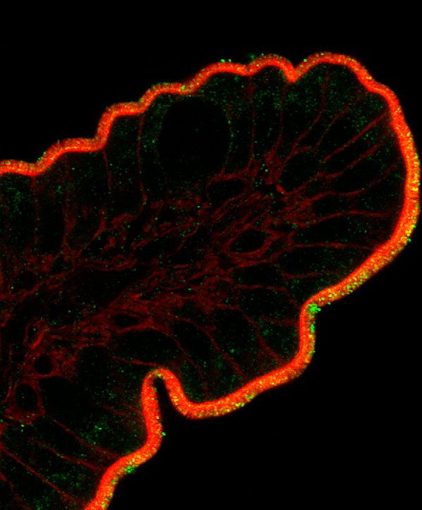
Fig. (1)

Expression of different astrocytic proteins following stroke. Increased expression of GFAP is a

hallmark of astrocytes activation, as is induction/re-expression of vimentin. Astrocytes normally

express glutamine synthetase (GS) and S100β, genes …

2.3. Forebrain Ischemia

Excitotoxic neuronal injury is a common mechanism in both acute and chronic

neurodegenerative diseases. It has long been appreciated that inhibition of astrocyte glutamate

uptake [34, 35], and more recently inhibition of astrocyte mitochondrial function [36], impairs

neuronal survival from excitotoxic injury. Brief forebrain ischemia is a model of the delayed

hippocampal neuronal loss seen in patients following cardiac arrest and resuscitation, and in part

involves excitotoxicity. Increased generation of reactive oxygen species (ROS) and

mitochondrial dysfunction in CA1 astrocytes contributes to ischemia-induced loss of GLT-1 and

ultimately to delayed death of CA1 neurons [15]. Our studies and those of other laboratories

have demonstrated that selective dysfunction of hippocampal CA1 subregion astrocytes, with

loss of glutamate transport activity and immunoreactivity for glutamate transporter 1 (GLT-1),

occurs at early reperfusion times, hours to days before the death of CA1 neurons [15, 37, 38].

The heterogeneous degeneration of astrocytic processes and mitochondria was tightly associated

with the appearance of disseminated selective neuronal necrosis and its maturation after

temporary ischemia [39]. By electronmicroscopy the same investigators [40] found that focal

infarction is exacerbated by temporary microvascular obstruction due to compression by swollen

astrocytic end-feet. However, hypoxia has multiple effects on astrocytes and their ability to

support neuronal viability [41]. For example, hypoxia induces astrocyte-dependent protection of

neurons following hypoxic preconditioning. Yet, hypoxia induces processes in astrocytes that

augment neuronal death in other situations, such as the coincidence of hypoxia with

inflammatory signaling.

3. REACTIVE ASTROGLIA: GOOD OR BAD AFTER STROKE?

The astrocyte response to ischemia has traditionally been viewed as detrimental to recovery, as

the astrocyte-rich glial scar has both physical and chemical inhibitory properties [42, 43]. As

components of the glial scar, astrocytes exhibit hypertrophied, interdigitated processes that form

a physical barrier. Astrocytes produce inhibitory molecules including chondroitin sulfate

proteoglycans (CSPGs) that contribute to chemical inhibition [44, 45]. In the acute setting,

astrocytic gap junctions may remain open following ischemia [46], allowing substances such as

proapoptotic factors to spread through the syncytium, thereby expanding the size of the infarct

[47]. As discussed below, astrocytes can also produce a variety of pro-inflammatory cytokines.

Many studies have shown that decreased astrogliosis often correlates with decreased infarct size.

Nonspecific inhibition of cell proliferation following ischemia using a cyclin kinase inhibitor

decreases astrocyte proliferation and results in improved functional recovery [48]. In addition,

treatment with alpha-melanocyte stimulating hormone [49], cysteinyl leukotriene receptor

antagonist [50], cliostazol [51], and caffeic acid [52] result in reduced infarct size accompanied

by a decrease in astrogliosis. Treadmill exercise [28] and acupuncture [53] are similarly

associated with improved outcome and reduced astrogliosis. Thus, results from several studies

suggest that treatments that decrease infarct size are often accompanied by attenuated astrocyte

response. Despite the frequent association of decreased astrogliosis with improved outcome, it is

difficult to determine cause and effect, since the extent of astrogliosis likely reflects the severity

of the injury, as well as influencing it.

In addition to their role in glial scar formation, astrocytes also respond to ischemia with functions

important for neuroprotection and repair. These include protecting spared tissue from further

damage [14], taking up excess glutamate as discussed above, rebuilding the blood brain barrier

[54, 55], and producing neurotrophic factors [10]. GFAP knockout mice exhibit larger lesions

than their wild-type littermates following focal ischemia [56], and mice lacking both GFAP and

vimentin have impaired astrocyte activation, decreased glutamate uptake abilities, and attenuated

PAI-1 expression after ischemia [57]. Application of astrocyte-conditioned media after transient

MCAO results in decreased infarct volume and regained blood-brain barrier function [58],

suggesting that factors released by astrocytes following ischemia are important for

neuroprotection.

Although few studies other than the use of animals lacking vimentin and GFAP have specifically

targeted astrocyte activation after ischemia, there is correlational evidence suggesting that

astrogliosis may be beneficial. Environmental enrichment, which results in reduced infarct size

and improved recovery following ischemia, also leads to increased astrocyte proliferation

[59, 60]. After focal ischemia, aged rats exhibit increased tissue damage and increased astrocyte

hypertrophy, but have decreased astrocyte proliferation compared to young rats [61]. Systemic

infusion of bone marrow stromal cells following MCAO increases gliogenesis and decreases

lesion size [62, 63]. In addition, administration of transforming growth factor α (TGFα), a known

mitogen for astrocytes [64], following MCAO leads to reduced infarct size and improved

functional recovery [65]. Furthermore, ischemic preconditioning that produces a neuroprotective

state leads to prolonged astrocyte expression of Hsp27 [66]. Finally, mice lacking connexin 43,

the gap junction connecting astrocyte networks that is needed for proper neurotransmitter and

potassium regulation, have increased infarcts following MCAO [67]. Thus, astrocytes have the

potential to be both detrimental and beneficial following ischemic insult, making them promising

targets for manipulation to improve outcome.

4. ASTROCYTE-MEDIATED INFLAMMATION AFTER STROKE: A DOUBLE-EDGED

SWORD

Inflammation plays both detrimental and beneficial roles in brain ischemia, depending upon the

timing and severity of the inflammation. Within minutes after injury, injured neurons in the core

and penumbra of the lesion and glial cells in the core produce pro-inflammatory mediators,

cytokines, and reactive oxygen species, which activate both astrocytes and microglia [68].

Activated astrocytes can produce the proinflammatory cytokines IL-6, TNFα, IL-1α and β,

interferon γ, and others [68–70]. High levels of these cytokines can be detrimental to ischemic

recovery [71–75] by directly inducing apoptosis of neuronal cells and/or increasing toxic nitric

oxide levels [76] and inhibiting neurogenesis [77]. Indeed, inactivation of astrocyte NfκB

signaling, shown to induce astrocyte production of pro-inflammatory cytokines [78], decreases

cytokine production and protects neurons after ischemic injury [79]. Hsp72 overexpression is

associated with lower NfκB activation and lower TNFα [80]. In addition to cytokines, reactive

astrocytes also produce chemokines following ischemia [81]. Chemokines upregulate adhesion

molecules in vascular endothelial cells, resulting in attraction of immune cells, which may

worsen ischemia-induced damage [82]. Overall, some aspects of the local inflammatory response

contribute to secondary injury to potentially viable tissue and lead to apoptotic and necrotic

neuronal cell death hours to days after injury [83], while other aspects are beneficial.

Although the potential benefits of inflammation after stroke have received relatively little

attention so far, indirect evidence suggests that some specific inflammatory reactions are

neuroprotective and neuroregenerative [84–91]. In addition to providing defense against the

invasion of pathogens, inflammation is also involved in clearing damaged tissue, and in

angiogenesis, tissue remodeling, and regeneration [89]. This is probably best studied in wound

healing, which is severely compromised if inflammation is inhibited [89, 91]. There is also

evidence suggesting that specific inflammatory factors can be protective in some circumstances.

IL-6, produced by astrocytes acutely after MCAO [69], is likely neuroprotective early after

ischemia [84]. Interestingly, ischemic preconditioning resulting in protection appears to be

dependent on TLR-4 signaling, and is accompanied by increased TNFα, NFκB, and COX-2

expression [90]. Indeed, in vitro work has shown that administration of TNFα in combination

with Hsp70 results in decreased expression of pro-apoptotic proteins following hypoxia [88].

Thus, it is important to consider these factors, along with timing, when trying to determine the

best strategy to reduce damage and improve recovery and regeneration.

5. ASTROCYTE SUPPORT OF NEURONS AFTER STROKE

5.1. Antioxidant Production

One hallmark of the cellular response to ischemia is a rapid, dramatic increase in damaging free

radicals, including nitric oxide (NO), superoxide, and peroxynitrite [92]. Nitric oxide synthetase

levels increase as soon as 10 minutes after induction of MCAO [93], followed by NO production

that persists for at least one week after MCAO [94]. Nitric oxide can cause cell death by

inducing the release of cytochrome-c from mitochondria, leading to apoptosis [95]. Nitric oxide

can also induce necrotic death [96]. Furthermore, the production of nitric oxide and other free

radicals can modify oxidative metabolism and impair ATP production [13, 19]. Changes in

mitochondrial properties can further limit oxidative metabolism [97]. Not surprisingly, several

studies have shown that antioxidant treatment enhances neuroprotection and recovery after

stroke [98–101].

The release of glutathione and SOD by astrocytes has been reported and is suggested to play an

important role in maintaining and enhancing neuronal survival, yet they are able to reduce

ascorbate for further neuronal antioxidant defense Fig. (2) [10, 102–106]. Interestingly, neurons

cocultured with astrocytes exhibit higher levels of glutathione compared with neurons cultured

alone [107]. Although astrocytes upregulate SOD after cerebral ischemia [108], they do not

appear to increase levels of glutathione in ischemic conditions [109]. It is unknown whether

ischemia alters astrocytic ascorbate levels, but osmotic swelling from ischemia results in

increased astrocyte release of ascorbate in vitro [110], suggesting that similar mechanisms may

occur in vivo.

Fig. (2)

Mechanisms of astrocyte support of neurons important in stroke. Antioxidant defense includes

release of glutathione and ascorbate. Regulation of extracellular levels of ions and neuro-

transmitters, especially K+ and glutamate, strongly influences neuronal …

Several treatments that attenuate ischemic injury result in increased glutathione levels [111, 112].

SOD converts superoxide into oxygen and hydrogen peroxide. Similar to glutathione, many

treatments that ameliorate stroke damage are accompanied by an increase in SOD [113, 114].

Furthermore, rodents overexpressing SOD1 have significantly smaller injuries after both focal

and global ischemia [115, 116], while mice with decreased SOD1 have larger infarcts [117].

Finally, ascorbate can also reduce oxidative stress [118]. Treatment with dehydroascorbic acid, a

blood-brain-barrier-permeable precursor to ascorbic acid, is protective after MCAO [119].

Dehydroascorbic acid is taken up by astrocytes and released as ascorbic acid [12], a process

increased by propofol [120], a treatment that can be protective after stroke [121]. In summary,

astrocytes are important producers of antioxidants in the normal CNS, and astrocyte production

of these molecules after stroke may enhance neuronal survival and protect astrocyte function.

5.2. Glutamate Regulation

Astrocytes are key players in the regulation of neuro-transmitters in the CNS. Astrocytes make

glutamine, the precursor for the neurotransmitters glutamate and GABA [122] Fig. (2). Astrocyte

production of neurotransmitter precursors is impaired after MCAO, and alterations in neuro-

transmitter levels occur throughout the brain following stroke, possibly contributing to neuronal

death [123, 124].

Astrocytes are primarily responsible for glutamate uptake in the normal brain using the astrocyte

specific glutamate transporters GLAST and GLT-1 (Fig. 2) [125–127], as excess glutamate leads

to cell death via excitotoxicity [128]. Glutamate transporter levels in astrocytes decrease acutely

following global ischemia [38, 129] and neonatal hypoxia-ischemia [130], most likely

exacerbating neuronal death as a result of glutamate-induced excitoxicity. Despite the therapeutic

potential of increasing astrocyte glutamate transport after stroke, few groups have explored this

possibility. Carnosine, shown to be protective after focal ischemia, may partially be effective

because it prevents loss of GLT-1 on astrocytes, resulting in attenuated excitotoxicity [131]. In a

more direct assessment of how post-ischemic astrocyte glutamate transporters contribute to

neuronal survival, our laboratory has shown that upregulation of GLT-1 on astrocytes using

ceftriaxone protects CA1 neurons after global ischemia [129], similar to its effects in focal

cerebral ischemia [132].

5.3. Potassium Uptake and Energy Metabolism

Astrocytes also regulate neuronal activation by extracellular potassium uptake [133] Fig. (2).

Neurons release potassium after activation, and increased extracellular potassium leads to

neuronal hyperexcitability [133], a phenomenon that occurs in ischemic conditions [134]. In

addition to regulating neuronal activation, proper maintenance of ion gradients, such as

potassium, is important in regulating cell volume in both normal and ischemic conditions

[135, 136]. Astrocytes increase potassium transporter activity in response to transient in

vitro ischemia [137]. Due to its effects on both neuronal activity and cell volume, increasing

astrocytic potassium uptake may be a possible therapeutic target for stroke.

Astrocytes are also integral to normal maintenance of neuronal metabolism. When astrocytes

take up extracellular glutamate as a result of neuronal activity, the Na+/ K+-ATPase, along with

AMPA signaling, triggers astrocyte uptake of glucose from the blood, as astrocytic endfeet

contact capillaries [138, 139]. This glucose is then made into lactate, a substrate for neuronal

energy, to further “fuel” active neurons [140] Fig. (2). As mentioned above, astrocytes produce

glutathione. In addition to its antioxidant properties, glutathione is needed for the conversion of

methylglyoxal, a toxic by-product of metabolism, into D-Lactate by glyoxalase 1 [141].

Although the role of astrocyte metabolism is relatively well-established in normal tissue, the

post-ischemic role of astrocyte metabolism maintenance is less clear [142]. After ischemia,

astrocytes upregulate glucose transporters in order to provide energy to stressed/dying neuronal

cells [143,144]. Ethyl pyruvate, a derivative of the energy substrate pyruvate, is neuroprotective

after stroke only when astrocytes are viable, suggesting that astrocytes are necessary for

improvement in post-ischemic energy metabolism [122].

6. NOVEL STRATEGIES TO IMPROVE THE NEURONAL SUPPORTIVE ROLE OF

ASTROCYTES

Although few studies have specifically targeted astrocytes for repair after stroke, there is some

evidence that this can be a successful strategy. Recent results indicate that induction of BDNF in

astrocytes by galectin-1 reduces neuronal apoptosis in ischemic boundary zone and improves

functional recovery [145]. In addition, protection by pyruvate against glutamate neurotoxicity is

mediated by astrocytes through a glutathione-dependent mechanism [146]. Our recent study

demonstrated that enhancing astrocyte resistance to ischemic stress by overexpressing protective

proteins or antioxidant enzyme results in improved survival of CA1 neurons following forebrain

ischemia Fig. (3) [16]. Two well-studied protective proteins, heat shock protein 72 (Hsp72) and

mitochondrial SOD, were genetically targeted for expression in astrocytes using the astrocyte-

specific human GFAP promoter. In both cases protection was accompanied by preservation of

the astrocytic glutamate transporter GLT-1, and reduced evidence of oxidative stress in the CA1

region [16]. Similarly, selective overexpression of excitatory amino acid transporter 2 (EAAT2)

in astrocytes enhances neuroprotection from moderate hypoxia-ischemia [147].

Fig. (3)

Targeted over-expression of Hsp72 in astrocytes reduces the vulnerability of CA1 neurons to

forebrain ischemia. Selective overexpression of Hsp72 in astrocytes by expressing it from the

astrocyte specific GFAP promoter was achieved by unilateral stereotaxic …

7. TRANSLATING INSIGHTS INTO PROTECTION INTO CLINICAL APPLICATIONS

Many factors have been identified that likely contribute to the failure in translation seen so far

with stroke therapies. Currently, the only approved stroke therapy is thrombolysis induced by

intravenous administration of recombinant tissue plasminogen activator [148]; however, because

of a short therapeutic time window, only a small fraction of patients benefit from this treatment.

Hypothermia is the only accepted acute treatment to reduce brain injury following cardiac arrest

and resuscitation [4]. Thus far many clinical trials have focused on treatments that would likely

be beneficial to neurons, with fewer studies focused on mechanisms that might benefit all cell

types or specifically targeting other cell types, such as astrocytes. Often the consequence of these

treatments on the astrocyte response is not considered. Several examples of past and ongoing

clinical trials are discussed below, with specific attention to how these treatments may alter

astrocyte response or viability.

Several clinical trials have targeted manipulation of the inflammatory response to ischemia, as

stroke patients with systemic inflammation exhibit poorer outcomes [149]. Although anti-

inflammatory therapy decreases infarct size and improves neurological sequelae in experimental

animal models of stroke [150], human trials with anti-neutrophil therapy have not shown a clear

benefit [151, 152]. In addition, recent clinical trials in which anti-CD11/18 antibodies were

administered to human subjects were unsuccessful [153]. Likewise, a double-blinded, placebo-

controlled clinical trial in which anti–ICAM-1 antibody was administered within 6 hours of

stroke symptoms showed disappointing results [151]. In understanding these results it is

important to recall that while experimental stroke is relatively homogeneous concerning size,

territory, and etiology, with more consistent inflammatory response, human stroke is extremely

heterogeneous [154], with different vascular territories and extents of injury. In addition, these

mediators are known to affect many organ systems beyond the central nervous system. Systemic

administration of anti-inflammatory agents may have exacerbated the relative state of

immunocompromise seen in stroke patients, thereby confounding the outcome. Furthermore,

inflammation and astrocyte response are likely closely connected. Although there is little

evidence for a direct relationship between neutrophils and astrocytes, it has been shown that

mice with a blunted inflammatory response exhibit increased loss of GFAP-positive astrocytes

after cortical stab injury [155]. Because astrocytic glial scar formation is important in protection

of spared tissue from further damage [156], it is possible that treatments that drastically attenuate

inflammation lead to a stunted astrocyte response that is deleterious to recovery.

Another drug that has advanced to clinical study is DP-b99, currently in phase III studies for

acute stroke. DP-b99 is a membrane active chelator derivative of the known calcium chelator,

BAPTA spell out [157]. A lipophilic chelator of calcium, zinc and copper ions, DP-b99

sequesters metal ions only within and in to cell membranes. This clinical trial is especially

attractive because sequestration of calcium, zinc, and copper are potentially beneficial not only

to neurons, but also to astrocytes. It has been shown in Alzheimer’s disease that beta amyloid

increases astrocyte calcium influx, which causes decreased glutathione levels [158]. Zinc

chloride is toxic to astrocytes as well as neurons in vitro [159]. Similarly, astrocytes exposed to

neocuprine exhibit increased copper influx and undergo apoptotic cell death [160]. Approaches

that benefit multiple cell types, including astrocytes, are more likely to be successful.

Other current ongoing clinical trials focus on neuroprotective agents for the purpose of aiding

neurological recovery after stroke. Minocycline (Phase I), edavarone (Phase IV), propanolol (a

β-blocker; phase II and III), and more recently arundic acid have been previously shown to be

protective and enhance neuronal survival in stroke [161–165], though by targeting different

mechanisms. Some additional completed and ongoing trials are summarized in Table 1.

Preclinical research needs to consider these clinical results, and assess effects on astrocytes as

well as neurons.

Table 1

Overview of Some Completed and Ongoing Clinical Trials for Stroke

Although anti-inflammatory strategies to diminish ischemic brain injury have failed thus far,

continued elucidation of the complex interactions involved in modulating the inflammatory

response may still enable novel therapeutic approaches that translate successfully into clinical

efficacy.

CONCLUSIONS

Traditionally, stroke research has focused on neurons and often ignored effects on glial cells. It is

increasingly evident that glia are vital to both normal CNS functioning and also play important

roles in neuropathological conditions. Although astrocytes form an inhibitory glial scar following

ischemia, they also perform functions necessary for neuronal survival and well-being, such as

maintaining low extracellular glutamate levels and providing antioxidant protection. Because

they have a great many functions, astrocytes are attractive candidates as therapeutic targets. By

striving to shift astrocytes towards a pro-reparative, neuronal-supportive phenotype following

stroke, future clinical therapies may well be more successful in protecting neurons from ischemic

damage and promoting repair.

ACKNOWLEDGEMENTS

This work was supported by NIH grants CM49831, N5053898, and NS014543 to RGG.

REFERENCES

1. Lloyd-Jones D, Adams RJ, Brown TM, Carnethon M, Dai S, De Simone G, Ferguson TB, Ford E, Furie K,

Gillespie C, Go A, Greenlund K, Haase N, Hailpern S, Ho PM, Howard V, Kissela B, Kittner S, Lackland D,

Lisabeth L, Marelli A, McDermott MM, Meigs J, Mozaffarian D, Mussolino M, Nichol G, Roger VL,

Rosamond W, Sacco R, Sorlie P, Roger VL, Thom T, Wasserthiel-Smoller S, Wong ND, Wylie-Rosett J.

Heart disease and stroke statistics–2010 update: a report from the American Heart

Association.Circulation. 2010;121:e46–e215. [PubMed]

2. Blakeley JO, Llinas RH. Thrombolytic therapy for acute ischemic stroke. J. Neurol.

Sci. 2007;261:55–62.[PubMed]

3. Marder VJ, Jahan R, Gruber T, Goyal A, Arora V. Thrombolysis with plasmin: implications for stroke

treatment. Stroke. 2010;41:S45–S49. [PMC free article] [PubMed]

4. Bernard SA, Gray TW, Buist MD, Jones BM, Silvester W, Gutteridge G, Smith K. Treatment of comatose

survivors of out-of-hospital cardiac arrest with induced hypothermia. N. Engl. J.

Med.2002;346:557–563. [PubMed]

5. Howells DW, Porritt MJ, Rewell SS, O’Collins V, Sena ES, van der Worp HB, Traystman RJ, Macleod MR.

Different strokes for different folks: the rich diversity of animal models of focal cerebral ischemia. J.

Cereb. Blood Flow Metab. 30:1412–1431. [PMC free article] [PubMed]

6. Kettenmann H, Ramson BR. Neuroglia. New York: Oxford University Press; 1995.

7. Nedergaard M, Ransom B, Goldman SA. New roles for astrocytes: redefining the functional

architecture of the brain. Trends Neurosci. 2003;26:523–530. [PubMed]

8. Sofroniew MV, Vinters HV. Astrocytes: biology and pathology. Acta

Neuropathol. 2010;119:7–35.[PMC free article] [PubMed]

9. Goldberg MP, Choi DW. Combined oxygen and glucose deprivation in cortical cell culture: calcium-

dependent and calcium-independent mechanisms of neuronal injury. J.

Neurosci. 1993;13:3510–3524.[PubMed]

10. Swanson RA, Ying W, Kauppinen TM. Astrocyte influences on ischemic neuronal death. Curr. Mol.

Med. 2004;4:193–205. [PubMed]

11. Dringen R, Hirrlinger J. Glutathione pathways in the brain. Biol. Chem. 2003;384:505–516. [PubMed]

12. Wilson JX. Antioxidant defense of the brain: a role for astrocytes. Can. J. Physiol.

Pharmacol.1997;75:1149–1163. [PubMed]

13. Rossi DJ, Brady JD, Mohr C. Astrocyte metabolism and signaling during brain ischemia. Nat.

Neurosci.2007;10:1377–1386. [PubMed]

14. Nedergaard M, Dirnagl U. Role of glial cells in cerebral ischemia. Glia. 2005;50:281–286. [PubMed]

15. Ouyang YB, Voloboueva LA, Xu LJ, Giffard RG. Selective dysfunction of hippocampal CA1 astrocytes

contributes to delayed neuronal damage after transient forebrain ischemia. J.

Neurosci.2007;27:4253–4260. [PMC free article] [PubMed]

16. Xu L, Emery JF, Ouyang YB, Voloboueva LA, Giffard RG. Astrocyte targeted overexpression of Hsp72

or SOD2 reduces neuronal vulnerability to forebrain ischemia. Glia. 2010;58:1042–1049.[PMC free

article] [PubMed]

17. Anderson MF, Blomstrand F, Blomstrand C, Eriksson PS, Nilsson M. Astrocytes and stroke:

networking for survival? Neurochem. Res. 2003;28:293–305. [PubMed]

18. Giffard RG, Swanson RA. Ischemia-induced programmed cell death in

astrocytes. Glia. 2005;50:299–306. [PubMed]

19. Almeida A, Delgado-Esteban M, Bolanos JP, Medina JM. Oxygen and glucose deprivation induces

mitochondrial dysfunction and oxidative stress in neurones but not in astrocytes in primary culture. J.

Neurochem. 2002;81:207–217. [PubMed]

20. Xu L, Sapolsky RM, Giffard RG. Differential sensitivity of murine astrocytes and neurons from

different brain regions to injury. Exp. Neurol. 2001;169:416–424. [PubMed]

21. Zhao G, Flavin MP. Differential sensitivity of rat hippocampal and cortical astrocytes to oxygen-

glucose deprivation injury. Neurosci. Lett. 2000;285:177–180. [PubMed]

22. Lukaszevicz AC, Sampaio N, Guegan C, Benchoua A, Couriaud C, Chevalier E, Sola B, Lacombe P,

Onteniente B. High sensitivity of protoplasmic cortical astroglia to focal ischemia. J. Cereb. Blood Flow

Metab. 2002;22:289–298. [PubMed]

23. Bondarenko A, Chesler M. Rapid astrocyte death induced by transient hypoxia, acidosis, and

extracellular ion shifts. Glia. 2001;34:134–142. [PubMed]

24. Giffard RG, Monyer H, Choi DW. Selective vulnerability of cultured cortical glia to injury by

extracellular acidosis. Brain Res. 1990;530:138–141. [PubMed]

25. Swanson RA, Farrell K, Stein BA. Astrocyte energetics, function, and death under conditions of

incomplete ischemia: a mechanism of glial death in the penumbra. Glia. 1997;21:142–153. [PubMed]

26. Tombaugh GC, Sapolsky RM. Mechanistic distinctions between excitotoxic and acidotic hippocampal

damage in an in vitro model of ischemia. J. Cereb. Blood Flow Metab. 1990;10:527–535. [PubMed]

27. Chen Y, Swanson RA. Astrocytes and brain injury. J. Cereb. Blood Flow

Metab. 2003;23:137–149.[PubMed]

28. Lee MH, Kim H, Kim SS, Lee TH, Lim BV, Chang HK, Jang MH, Shin MC, Shin MS, Kim CJ. Treadmill

exercise suppresses ischemia-induced increment in apoptosis and cell proliferation in hippocampal

dentate gyrus of gerbils. Life Sci. 2003;73:2455–2465. [PubMed]

29. Li Y, Chopp M, Zhang ZG, Zhang RL. Expression of glial fibrillary acidic protein in areas of focal

cerebral ischemia accompanies neuronal expression of 72-kDa heat shock protein. J. Neurol.

Sci.1995;128:134–142. [PubMed]

30. Schroeter M, Schiene K, Kraemer M, Hagemann G, Weigel H, Eysel UT, Witte OW, Stoll G. Astroglial

responses in photochemically induced focal ischemia of the rat cortex. Exp Brain

Res. 1995;106:1–6.[PubMed]

31. Van Beek J, Chan P, Bernaudin M, Petit E, MacKenzie ET, Fontaine M. Glial responses, clusterin, and

complement in permanent focal cerebral ischemia in the mouse. Glia. 2000;31:39–50. [PubMed]

32. Yamashita K, Vogel P, Fritze K, Back T, Hossmann KA, Wiessner C. Monitoring the temporal and

spatial activation pattern of astrocytes in focal cerebral ischemia using in situ hybridization to GFAP

mRNA: comparison with sgp-2 and hsp70 mRNA and the effect of glutamate receptor antagonists. Brain

Res.1996;735:285–297. [PubMed]

33. Liu D, Smith CL, Barone FC, Ellison JA, Lysko PG, Li K, Simpson IA. Astrocytic demise precedes delayed

neuronal death in focal ischemic rat brain. Brain Res. Mol. Brain Res. 1999;68:29–41. [PubMed]

34. Dugan LL, Bruno VM, Amagasu SM, Giffard RG. Glia modulate the response of murine cortical

neurons to excitotoxicity: glia exacerbate AMPA neurotoxicity. J.

Neurosci. 1995;15:4545–4555. [PubMed]

35. Rosenberg PA, Aizenman E. Hundred-fold increase in neuronal vulnerability to glutamate toxicity in

astrocyte-poor cultures of rat cerebral cortex. Neurosci. Lett. 1989;103:162–168. [PubMed]

36. Voloboueva LA, Suh SW, Swanson RA, Giffard RG. Inhibition of mitochondrial function in astrocytes:

implications for neuroprotection. J. Neurochem. 2007;102:1383–1394. [PMC free article] [PubMed]

37. Chen JC, Hsu-Chou H, Lu JL, Chiang YC, Huang HM, Wang HL, Wu T, Liao JJ, Yeh TS. Down-regulation

of the glial glutamate transporter GLT-1 in rat hippocampus and striatum and its modulation by a group

III metabotropic glutamate receptor antagonist following transient global forebrain ischemia. Neuro-

pharmacology. 2005;49:703–714. [PubMed]

38. Yeh TH, Hwang HM, Chen JJ, Wu T, Li AH, Wang HL. Glutamate transporter function of rat

hippocampal astrocytes is impaired following the global ischemia. Neurobiol.

Dis. 2005;18:476–483.[PubMed]

39. Ito U, Hakamata Y, Kawakami E, Oyanagi K. Degeneration of astrocytic processes and their

mitochondria in cerebral cortical regions peripheral to the cortical infarction: heterogeneity of their

disintegration is closely associated with disseminated selective neuronal necrosis and maturation of

injury.Stroke. 2009;40:2173–2181. [PubMed]

40. Ito U, Hakamata Y, Kawakami E, Oyanagi K. Temporary focal cerebral ischemia results in swollen

astrocytic end-feet that compress microvessels and lead to focal cortical infarction. J. Cereb. Blood Flow

Metab. 2010 [PMC free article] [PubMed]

41. Vangeison G, Rempe DA. The Janus-faced effects of hypoxia on astrocyte

function. Neuroscientist.2009;15:579–588. [PMC free article] [PubMed]

Read More

L-Theanine and Caffeine in Combination Affect Human Cognition as Evidenced by Oscillatory alpha- Band Activity and Attention Task Performance1–3

L-Theanine and Caffeine in Combination Affect

Human Cognition as Evidenced by Oscillatory alpha-

Band Activity and Attention Task Performance1–3

1. Simon P. Kelly,

2. Manuel Gomez-Ramirez,

3. Jennifer L. Montesi, and

4. John J. Foxe*

+Author Affiliations

1. Cognitive Neurophysiology Laboratory, Nathan S. Kline Institute for Psychiatric Research,

Program in Cognitive Neuroscience and Schizophrenia, Orangeburg, NY 10962 and Program

in Cognitive Neuroscience, Department of Psychology, City College of the City University of

New York, New York, NY 10031

1. ↵*To whom correspondence should be addressed. E-mail: [email protected].


Next Section

Abstract

Recent neuropharmacological research has suggested that certain constituents of tea may have modulatory

effects on brain state. The bulk of this research has focused on either L-theanine or caffeine ingested alone

(mostly the latter) and has been limited to behavioral testing, subjective rating, or neurophysiological

assessments during resting. Here, we investigated the effects of both L-theanine and caffeine, ingested

separately or together, on behavioral and electrophysiological indices of tonic (background) and phasic (event-

related) visuospatial attentional deployment. Subjects underwent 4 d of testing, ingesting either placebo, 100

mg of L-theanine, 50 mg of caffeine, or these treatments combined. The task involved cued shifts of attention to

the left or right visual hemifield in anticipation of an imperative stimulus requiring discrimination. In addition to

behavioral measures, we examined overall, tonic attentional focus as well as phasic, cue-dependent

anticipatory attentional biasing, as indexed by scalp-recorded alpha-band (8–14 Hz) activity. We found an

increase in hit rate and target discriminability (d′) for the combined treatment relative to placebo, and an

increase in d′ but not hit rate for caffeine alone, whereas no effects were detected for L-theanine alone.

Electrophysiological results did not show increased differential biasing in phasic alpha across hemifields but

showed lower overall tonic alpha power in the combined treatment, similar to previous findings at a larger

dosage of L-theanine alone. This may signify a more generalized tonic deployment of attentional resources to

the visual modality and may underlie the facilitated behavioral performance on the combined ingestion of these

2 major constituents of tea.

Previous SectionNext Section

Introduction

In recent years, several potential health benefits of drinking tea (Camellia sinensis) have come to light through

systematic study of the effects of its constituent compounds (1,2). Although anecdotal evidence abounds, the

psychological and neurophysiological effects of tea have received relatively little experimental investigation and

thus remain unclear. Popular claims have centered on generalized state changes such as the reduction of

stress and induction of relaxed wakefulness. Psychopharmacological studies have indeed demonstrated mood

effects that support these claims and have further shown that tea affects elements of cognition (3,4). Although

caffeine (1,3,7-trimethylxanthine) is by far the constituent most studied, with findings of increased alertness and

speeded reaction time (RT)4 predominant (5,6), there exists evidence that caffeine alone cannot fully account

for the positive effects of tea drinking. Tea has been shown to raise skin temperature to a higher level (7), to

increase critical flicker fusion threshold (4), and to reduce physiological stress responses and increase

relaxation ratings (8) when compared with coffee or other control beverages matched for caffeine level.

L-Theanine (γ-glutamylethylamide), a unique amino acid present almost exclusively in the tea plant, has

recently received research interest in the neuroscience community with findings of neuroprotective effects [see

Kakuda (9)] and mood effects indexed both by subjective self-reports (10) and via psychological and

physiological responses to stress (11). Using electroencephalographic (EEG) recordings in humans, Kobayashi

et al. (12) and Juneja et al. (13) reported that activity within the alpha frequency band (8–14 Hz) increased in

reaction to L-theanine ingestion when measured during a state of rest. This was of interest to the attention

community, as the alpha rhythm has long been known to be sensitive to overall attentional states (i.e., intensity

aspects such as arousal) (14) and, further, is involved in the biasing of selective attention (15,16). In

intersensory attention tasks, where the relevant modality is cued ∼1 s before a compound audiovisual target

stimulus, parieto-occipital alpha power in the intervening period is increased for attend-visual trials relative to

attend-auditory trials (15,17). In Gomez-Ramirez et al. (18), this differential effect of cue information on

anticipatory alpha amplitude was found to be larger on ingestion of 250 mg of L-theanine relative to placebo. In

addition, tonic (background) alpha amplitude was relatively decreased for L-theanine, in apparent contradiction

to the findings of Juneja et al. (13). In a follow-up study, we tested whether an analogous alpha-mediated

attention effect seen in visuospatial attention tasks (16,19–22) is also affected by L-theanine ingestion (M.

Gomez-Ramirez, S. P. Kelly, J. L. Montesi, and J. J. Foxe, unpublished results). L-Theanine, at a dosage of

250 mg, was not found to increase the differential effect of attention. However, in a replication of the previous

intersensory attention study (18), overall tonic alpha was greatly reduced on L-theanine.

An immediate question, given this replication, is whether this tonic alpha reduction occurs at lower dosages

of L-theanine, closer to the amount ingested through a typical serving of tea. In the present study, we

administered a lower dosage of 100 mg to address this. Also of critical interest is whether the ingestion of

caffeine, another major component of tea, exerts behavioral and/or neurophysiological effects during such a

demanding visuospatial attention task, when ingested alone or when ingested together with L-theanine. Here

we present data from a 4-d experiment using a balanced repeated-measures design, with subjects receiving

either placebo (P), L-theanine alone (T), caffeine alone (C), or the combination of L-theanine plus caffeine

(T+C) on each day. We assessed effects of treatment with regard to basic behavioral measures of RT and

accuracy [including the so-called discriminability index (d′), which is independent of individual detection criteria],

and in relation to both tonic and phasic attentional processes as indexed by alpha power.

Previous SectionNext Section

Methods

Participants.

Sixteen (5 female) neurologically normal paid volunteers, aged between 21 and 40 y (mean 27.5 y),

participated in the study. All subjects provided written informed consent, and the Institutional Review Board of

the Nathan S. Kline Institute for Psychiatric Research approved the experimental procedures. All subjects

reported normal or corrected-to-normal vision. Four subjects were left-handed. The mean habitual tea

consumption across the subjects was 3.7 cups/wk, and for coffee, 3.8 cups/wk (∼250 mL/cup). Subjects arrived

at the laboratory in the morning between 0900 and 1200 h, having abstained from all caffeinated beverages for

the previous 24 h.

Treatment.

The timing of treatment administration relative to testing was based on published reports of amino acid

concentration and plasma concentration changes over time.L-Theanine concentration has been found to

increase significantly within 1 h after administration in rats, to continue to increase gradually up to 5 h, and to

decrease thereafter, with complete disappearance evident after 24 h (23). Peak plasma caffeine concentration

is reached between 15 and 120 min postingestion in humans, with a variable half-life typically between 2.5 and

4.5 h (5). Accordingly, participants abstained from consuming caffeine for at least 24 h before testing and

began experimental task runs 30 min after ingestion of any given treatment. Subjects underwent 4 d of testing,

ingesting either placebo, 100 mg of L-theanine, 50 mg of caffeine, or these treatments combined. Subjects

were uninformed of the treatment, which was served in 100 mL of water, with the placebo treatment consisting

only of water. Both theanine and caffeine are tasteless in water solution.

Stimuli and task.

Subjects were seated 150 cm from a CRT monitor and were instructed to maintain fixation on a central cross

(white on midgray background) at all times. Each trial began with a centrally presented arrow cue (“S1”) of 100-

ms duration, with equal probability pointing leftward or rightward toward 1 of 2 bilateral locations centered at a

horizontal distance of 4.2° from the fixation cross and 1.2° above the horizontal meridian. Each location was

marked by 4 dots outlining a 2.4° × 2.4° square. The cue consisted of a circle of 1° diameter with an embedded

arrow, designed to minimize any sensory effects related to physical differences between the left and right cues.

The colors of the arrow and circle were red on green for half of the blocks of recording and green on red for the

other half, with the order counterbalanced across subjects and days of testing. Red and green values were

precalibrated for each subject to be approximately isoluminant by flicker photometry. Then, 933 ms after cue

onset, a second imperative stimulus (“S2”) was presented at the left or right marked location (valid or invalid

with respect to cue direction) with equal probability. The S2s (100 ms duration) consisted of either a white × or

+ (0.75° × 0.75°) embedded in a circular array of 8 small circles such that the overall stimulus diameter was

1.95°. The target stimulus was chosen randomly at the beginning of each experimental run of ∼4.5 min, and

thereafter standard and target stimuli were equally likely on each trial. Subjects were instructed to shift their

attention covertly to the location indicated by the cue, to respond by pressing a mouse button with the index

finger of the right hand when a target S2 appeared on that side, and to ignore stimuli appearing on the invalid

side entirely. Trials were separated by a 1633-ms interval. A total of 100 trials were presented per run. Subjects

completed 20 runs on each day of testing.

Data acquisition.

Continuous EEG data, digitized at 512 Hz, were acquired from 164 scalp electrodes and 4 electro-oculographic

(EOG) electrodes with a pass-band of 0.05–100 Hz. Off-line, the data were low-pass filtered up to 45 Hz and

rereferenced to the nasion. Noisy channels, identified by taking the SD of amplitude over the entire run (from

first to last stimulus presented) and checking whether it is >50% greater than that of at least 3 of the 6 closest

surrounding channels, were interpolated. Horizontal EOG data were recorded using 2 electrodes placed at the

outer canthi of the eyes, allowing measurement of eye movements during testing. Based on a calibrated

mapping of EOG amplitude to visual angle, trials were rejected off-line if eye gaze deviated by >0.5° during the

cue-target interval.

Behavioral data analysis.

We employed a d′ as our principal performance metric, taking into account the accuracy of responding on

nontargets as well as targets and controlling for individual differences in detection criteria. The value of d′ was

derived from the hit rate (proportion of all valid targets detected) and false alarm rate (proportion of all valid

nontargets incorrectly responded to), calculated only from trials containing no eye movements or artifacts.

Ceiling effects on hit rate were corrected in the standard way by assuming 0.5 misses, and similarly, a floor

effect of zero false alarms was corrected to 0.5. RT was measured as the time (in milliseconds) from the point

of S2 onset at which the mouse button was correctly pressed in response to valid target trials.

To control for the potential confound of practice effects on the behavioral data, the order of treatments across

the 4 d of testing was fully counterbalanced across subjects. This is a standard procedure and ensures

unbiased comparison across conditions. However, in the case of the present data, the variance in behavioral

measures arising from the day of testing (order effect) far superseded that arising from treatment. Thus, a

normalization of these measures was necessary to remove the variance caused by practice, and this was

carried out by transforming each data point to a z-score with respect to the mean and SD of all scores

measured on that day (d 1, d 2, d 3, d 4). Because the distribution of scores for each day contains an equal

number of data points from each treatment, it cannot result in any bias for treatment but, rather, optimizes

statistical power to test for treatment effects.

Electrophysiological data analysis.

EEG data were epoched from −300 ms before to 1100 ms after cue onset and baseline-corrected relative to

the interval −100 to 0 ms, with an artifact rejection threshold of ±100 μV applied. Mean alpha amplitude was

calculated using the temporal spectral evolution (TSE) technique (15). TSE is carried out simply by filtering

each epoch with a passband of 8–14 Hz, rectifying, then averaging across trials. The averaged TSE waveforms

were then smoothed by averaging data points within a sliding 100-ms window.

The first analysis concerned tonic (background) alpha amplitude, which was found to decrease on L-theanine in

our previous 2 studies (18, M. Gomez-Ramirez, S. P. Kelly, J. L. Montesi, and J. J. Foxe, unpublished results).

Tonic alpha was measured as the integrated TSE amplitude within the baseline period −200 to 0 before the cue

stimulus, regardless of the direction of attentional deployment (i.e., to the left or right hemifield). The dependent

measure was computed as the baseline alpha amplitude averaged across 6 electrodes, chosen on the basis of

the grand-average scalp distribution of alpha amplitude, collapsed across the 4 d.

In a second analysis, we tested lateralized, anticipatory alpha amplitude for effects of attention and possible

interactions with treatment. We normalized alpha amplitude relative to baseline by dividing the TSE amplitude

by the mean amplitude within the baseline interval (−200 to 0) and log-transforming, making the measure

equivalent to a percentage change from baseline. This narrows down the analysis to attention-related

differential activity, independent of tonic effects. The anticipatory alpha dependent measure was computed as

the integrated TSE amplitude over the postcue interval 500 to 900 ms, ending just before the S2. Amplitude

was averaged across 6 electrodes over each hemisphere, determined based on grand-average difference

topographies (cue-left minus cue-right) collapsed across the 4 d.

Statistical methods.

A 4-d balanced repeated-measures design was employed, with subjects receiving 1 of the 4 treatments

(including placebo) on each day in counterbalanced order. SPSS for Windows (version 12.0) was used for all

statistical analyses. Tests were conducted with an α level of 0.05 unless otherwise stated. In the analysis of

behavioral data, we tested specifically for improvements in performance as a result of any of the 3 treatments.

Thus, 1-tailed, paired t-tests (df = 15) were conducted between the placebo condition and each of the 3

treatments for hit rate, RT, and d′ measures. Because 3 t-tests were performed including the same placebo

data, we applied a Bonferroni-corrected α-level of 0.016 here.

To test for effects of tonic alpha amplitude, a 1-way ANOVA was carried out with the factor of treatment having

the levels P, T, C, and T+C. Follow-up protected ttests were then conducted to unpack significant differences

existing between each of the T, C, and T+C conditions and the P condition. Further post hoc paired

comparisons among the 4 treatment conditions were conducted as appropriate through additional t-tests.

To test for effects on pretarget alpha amplitude a 4 × 2 × 2 ANOVA was carried out with factors of treatment (P,

T, C, T+C), attention (cue-left, cue-right), and hemisphere (left, right). To unpack a potential 3-way interaction,

we reduced the alpha cueing effect (typically seen as a hemisphere × attention interaction) to a single measure

by adding the differential over the 2 hemispheres, i.e., subtracting cue-right from cue-left on the left

hemisphere, subtracting cue-left from cue-right on the right hemisphere, and summing these 2 values. Thus

reduced, testing of treatment effects on the alpha cueing effect, as found in the analogous intersensory study of

Gomez-Ramirez et al. (18), could be done via paired t-tests comparing each of the 3 treatments T, C, and T+C

against P.

Previous SectionNext Section

Results

Behavioral performance.

Behavioral performance was significantly improved on the combined treatment (T+C) in terms of hit rate (P <

0.016) and d′ (P < 0.002). There was also a significant improvement in d′ on C compared with P (P < 0.016),

but not in hit rate. There were no significant effects of L-theanine, and no effects of any of the 3 treatments on

RT (Fig. 1).

 Download as PowerPoint Slide

FIGURE 1

Mean hit rate (proportion of targets detected) (upper panel), mean d′ (middle panel), and mean RT (lower

panel) when subjects ingested placebo (P),L-theanine (T), caffeine (C), or these treatments combined (T+C).

Values are means (n = 16). Asterisks indicate difference from P: *P < 0.05, **P < 0.01).

Electrophysiology.

There was a significant effect of treatment on tonic alpha amplitude (P < 0.02). Follow-up t-tests revealed that

alpha was significantly lower for T+C than P (P < 0.02). P did not differ from either T or C (see Fig. 2). Tonic

alpha differed between T+C and T (P < 0.005) but not between T+C and C.

 Download as PowerPoint Slide

FIGURE 2

TSE waveforms at midline electrodes from which the baseline tonic alpha measure was derived (upper panel).

Cue-left and cue-right trials are collapsed. Integrated amplitude over the baseline period for each treatment,

with significant difference from placebo marked with an asterisk (lower panel). The electrodes from which tonic

alpha measures were derived are marked on the 168-channel montage.

The typical alpha cueing effect was readily apparent in both the nonnormalized alpha amplitude waveforms and

normalized pretarget measures (Fig. 3) for each treatment day. A strong attention × hemisphere interaction

(P < 0.0005) was found on the pretarget anticipatory alpha measures as expected, reflecting the typically

observed alpha-mediated cueing effect. In addition, there was a significant 3-way interaction among treatment,

attention, and hemisphere (P < 0.05). When we reduced the alpha cueing effect to a single metric as described

above, the effect was smaller on C than P (P < 0.02) but did not differ for the T or T+C conditions.

 Download as PowerPoint Slide

FIGURE 3

(Upper panel) TSE waveforms over left and right hemispheres, with cue-left (solid) and cue-right (dashed)

superimposed, collapsed across treatment. The overall alpha-mediated spatial cueing effect is highlighted.

Electrode sites for cueing effect measurement are marked on the montage. (Lower panel) Normalized alpha

measures forming the dependent variable in tests for effects of treatment on the alpha cueing effect (P,

placebo; T, L-theanine; C, caffeine; T+C, combined).

Previous SectionNext Section

Discussion

This study was aimed at extending our knowledge of the effects of compounds contained in tea on the

cognitive function of attention. Testing relatively low-dosage treatments of L-theanine alone (100 mg), caffeine

alone (50 mg), and their combination, we observed an interesting pattern of effects for both behavioral and

electrophysiological measures. Whereas no behavioral effects on hit rate were apparent for either treatment

alone at the low dosages tested here, when both L-theanine and caffeine were ingested together, hit rate

underwent an enhancement of ∼3%. In terms of d′, improvements were seen for both caffeine alone and L-

theanine plus caffeine, the latter having a larger effect size (0.55 vs. 0.42 calculated as Cohen’s d). Given the

absence of any difference in hit rate for caffeine, the d′ effect must result from subjects making fewer false

alarms on caffeine.

Tonic alpha amplitude was not found to decrease significantly on the lower dosage of L-theanine. This indicates

that the effect is dose dependent because a drop was seen in both of our previous studies using a 250-mg

dosage (18, M. Gomez-Ramirez, S. P. Kelly, J. L. Montesi, and J. J. Foxe, unpublished results). There was,

however, a significant decrease in tonic alpha for the combined treatment. That this decrease marks a synergy

between the 2 compounds is suggested by the numerical difference in the alpha decrease caused by L-

theanine with and without caffeine (Fig. 2). That is, it seems unlikely that the greater decrease on the combined

treatment is simply a linear sum of the decreases from each compound alone. Because only single dosages of

each compound were tested, however, a fair degree of caution is appropriate in the interpretation of synergy at

this juncture. This study marks the third finding of decreased alpha as a result of L-theanine ingestion (albeit a

partial cause here) to date, demonstrating the reliability of the effect. At this point, the question of whether it

translates to an improved functional brain state requires serious consideration. Should the finding of a decrease

be received with positive connotations for health and/or mental capabilities?

In the 80 y since the discovery of alpha waves (24), alpha has been measured in almost any experimental

situation and human population, with significant effects abounding, but with a complicated picture and quite

disparate theoretical frameworks arising (25–26). A consistent principle appears to be that stronger alpha infers

positive functioning across individuals (27,28), whereas phasic changes within individuals reflect immediate

stimulus processing and anticipatory enhancement and/or suppression, with a greater retinotopically specific

decrease in alpha being predictive of better detection performance (21). The tonic depression of alpha during

task performance over the day of testing, as observed here, is neither an individual trait nor a phasic event-

related response but a lasting, tonic treatment effect, making it difficult to draw comparisons with such previous

studies. The finding of increased alpha on ingestion of theanine has previously been taken to indicate

increased relaxation without increased drowsiness (13). But this qualification appears tenuous in light of other

observations of treatment-related increased alpha, e.g., during marihuana-induced euphoria (29). Can “good”

and “bad” really be ascribed to increases and decreases in alpha, in whatever direction? Certainly, that this

treatment-related decrease in tonic alpha does not have negative implications is suggested, if not already by

the fact that tea has been keenly, routinely consumed for centuries, by the concomitant facilitation in behavioral

performance found here in terms of both hit rate and d′.

Previous studies have reported a drop in absolute alpha power during resting with eyes open on ingestion of

caffeine at higher dosages, e.g., 200 mg (30) and 400 mg (31). Although alpha amplitude was numerically

lower on 50 mg of caffeine alone here, this did not reach significance (P = 0.18). From this, it is clear that alpha

effects of both L-theanine and caffeine are dose dependent, demonstrating that full characterization of dose-

response functions in future studies is called for.

Evidence of a synergistic relationship between L-theanine and caffeine has been presented in recent

behavioral studies. Parnell et al. (32) reported improved speed and accuracy on an attention-switching task at

60 min and reduced susceptibility to distracting information during a memory task at both 60 and 90 min

following ingestion of a combination of L-theanine and caffeine in the same dosages as used here. Haskell et

al. (33) administered a large battery of cognitive tests before and after consumption of a drink containing either

placebo, 250 mg of L-theanine, 150 mg of caffeine, or their combination. These authors found improvements in

simple and numeric working memory RT, sentence verification accuracy, and alertness ratings for the

combined treatment but not for either treatment alone. Using a similar crossover design but with a greater

dosage of caffeine (250 mg) than L-theanine (200 mg), Rogers et al. (34) found that L-theanine tended to

counteract the caffeine-induced rise in blood pressure but did not interact with caffeine-induced increases in

either alertness or “jitteriness” on state anxiety scales. Although the measures examined in these investigations

and our study are quite distinct in nature, an emerging possibility is that the presence of synergistic effects

closely hinges on dosages. That is, it may be that theanine was not effective in augmenting the caffeine-

induced effects in Rogers et al. (34) because these were present at a saturated level. In the present study,

lower dosages were used, and a significant drop in tonic alpha was observed for L-theanine and caffeine

ingested together but not for either L-theanine or caffeine when ingested alone. However, the absence of a

significant difference between the caffeine-alone and combined treatments calls for caution in making strong

claims of synergy at this point.

Similar to our previous visuospatial attention study (M. Gomez-Ramirez, S. P. Kelly, J. L. Montesi, and J. J.

Foxe, unpublished results), we did not find any change in the alpha differential cueing effect for the L-theanine-

alone treatment. However, it is interesting that the cueing effect was found to be smaller on caffeine alone but

not for the combined treatment. This result was unexpected and thus will bear replication and further

investigation. For now, it appears that within visual space, attentional biasing as indexed by alpha amplitude is

not affected by L-theanine. In contrast, the cued biasing of attention between sensory modalities does appear

to be affected (18). A tentative interpretation of the current pattern of results is thatL-theanine works to enhance

the tonic apportionment of attentional resources to the visual modality and does so to a significant degree when

a large dosage is ingested by itself or in combination with caffeine when a smaller dosage is ingested.

Other articles in this supplement include references (35–44).

Previous SectionNext Section

Footnotes

 ↵1 Published in a supplement to The Journal of Nutrition. Presented at the conference “Fourth International

Scientific Symposium on Tea and Human Health,” held in Washington, DC at the U.S. Department of

Agriculture on September 18, 2007. The conference was organized by the Tea Council of the U.S.A. and was

cosponsored by the American Cancer Society, the American College of Nutrition, the American Medical

Women’s Association, the American Society for Nutrition, and the Linus Pauling Institute. Its contents are solely

the responsibility of the authors and do not necessarily represent the official views of the Tea Council of the

U.S.A. or the cosponsoring organizations. Supplement coordinators for the supplement publication were

Lenore Arab, University of California, Los Angeles, CA and Jeffrey Blumberg, Tufts University, Boston, MA.

Supplement coordinator disclosure: L. Arab and J. Blumberg received honorarium and travel support from the

Tea Council of the U.S.A. for cochairing the Fourth International Scientific Symposium on Tea and Human

Health and for editorial services provided for this supplement publication; they also serve as members of the

Scientific Advisory Panel of the Tea Council of the U.S.A.

 ↵2 Author disclosures: S. P. Kelly, M. Gomez-Ramirez, and J. L. Montesi, no conflicts of interest; J. J. Foxe

received an honorarium and travel support from the Tea Council of the U.S.A. for speaking at the Fourth

International Scientific Symposium on Tea and Human Health and for preparing this manuscript for publication.

 ↵3 Supported by a grant from the Lipton Institute of Tea in association with Unilever Beverages Global

Technology Centre in Colworth House, Sharnbrook, UK.

 ↵4 Abbreviations used: C, caffeine-alone condition; d′, discriminability index; EEG, electroencephalographic;

EOG, electro-oculographic; P, placebo condition; RT, reaction time; T, theanine-alone condition; T+C,

combined condition; TSE, temporal spectral evolution.

Previous Section


LITERATURE CITED

1. 1.↵

Yang CS, Landau JM. Effects of tea consumption on nutrition and health. J Nutr. 2000;130:2409–12.


Abstract/FREE Full Text

2. 2.↵

Blumberg J. Introduction to the Proceedings of the Third International Scientific Symposium on Tea and

Human Health: role of flavonoids in the diet. J Nutr. 2003;133:3244S–6S.


FREE Full Text

3. 3.↵

Quinlan PT, Lane J, Moore KL, Aspen J, Rycroft JA, O’Brien DC. The acute physiological and mood effects of

tea and coffee: the role of caffeine level.Pharmacol Biochem Behav. 2000;66:19–28.


CrossRefMedline

4. 4.↵

Hindmarch I, Rigney U, Stanley N, Quinlan P, Rycroft J, Lane J. A naturalistic investigation of the effects of

day-long consumption of tea, coffee and water on alertness, sleep onset and sleep

quality. Psychopharmacology (Berl).2000;149:203–16.


CrossRefMedline

5. 5.↵

Fredholm BB, Battig K, Holmen J, Nehlig A, Zvartau EE. Actions of caffeine in the brain with special reference

to factors that contribute to its widespread use.Pharmacol Rev. 1999;51:83–133.


FREE Full Text

6. 6.↵

Smith A. Effects of caffeine on human behaviour. Food Chem Toxicol.2002;40:1243–55.


CrossRefMedline

7. 7.↵

Quinlan P, Lane J, Aspinall L. Effects of hot tea, coffee and water ingestion on physiological responses and

mood: The role of caffeine, water and beverage type.Psychopharmacology (Berl). 1997;134:164–73.


CrossRefMedline

8. 8.↵

Steptoe A, Gibson EL, Vounonvirta R, Williams ED, Hamer M, Rycroft JA, Erusalimsky JD, Wardle J. The

effects of tea on psychophysiological stress responsivity and post-stress recovery: a randomised double-blind

trial.Psychopharmacology (Berl). 2007;190:81–9.


CrossRefMedline

9. 9.↵

Kakuda T. Neuroprotective effects of the green tea components L-theanine and catechins. Biol Pharm

Bull. 2002;25:1513–8.


CrossRefMedline

10. 10.↵

Lu K, Gray MA, Oliver C, Liley DT, Harrison BJ, Bartholomeusz CF, Phan KL, Nathan PJ. The acute effects

of L-theanine in comparison with alprazolam on anticipatory anxiety in humans. Hum

Psychopharmacol. 2004;19:457–65.

CrossRefMedline

11. 11.↵

Kimura K, Ozeki M, Juneja LR, Ohira H. L-Theanine reduces psychological and physiological stress

responses. Biol Psychol. 2007;74:39–45.


CrossRefMedline

12. 12.↵

Kobayashi K, Nagata Y, Aloi N, Juneja LR, Kim M, Yamamoto T, Sugimoto S. Effects of L-theanine on the

release of α-brain waves in human volunteers. Nihon Nogeikagaku Kaishi. 1998;72:153–7.

13. 13.↵

Juneja LR, Chu DC, Okubo T, Nagato Y, Yokogoshi H. L-Theanine—a unique amino acid of green tea and its

relaxation effect in humans. Trends Food Sci Technol. 1999;10:199–204.


CrossRef

14. 14.↵

Adrian ED, Matthews BHC. The Berger Rhythm: Potential changes in the occipital lobes in

man. Brain. 1934;57:355–85.


FREE Full Text

15. 15.↵

Foxe JJ, Simpson GV, Ahlfors SP. Parieto-occipital ∼10 Hz activity reflects anticipatory state of visual

attention mechanisms. Neuroreport. 1998;9:3929–33.

CrossRefMedline

16. 16.↵

Kelly SP, Lalor EC, Reilly RB, Foxe JJ. Increases in alpha oscillatory power reflect an active retinotopic

mechanism for distracter suppression during sustained visuospatial attention. J

Neurophysiol. 2006;95:3844–51.

Abstract/FREE Full Text

17. 17.↵

Fu KM, Foxe JJ, Murray MM, Higgins BA, Javitt DC, Schroeder CE. Attention-dependent suppression of

distracter visual input can be cross-modally cued as indexed by anticipatory parieto-occipital alpha-band

oscillations. Brain Res Cogn Brain Res. 2001;12:145–52.


CrossRefMedline

18. 18.↵

Gomez-Ramirez M, Higgins BA, Rycroft JA, Owen GN, Mahoney J, Shpaner M, Foxe JJ. The deployment of

intersensory selective attention: a high-density electrical mapping study of the effects of theanine. Clin

Neuropharmacol.2007;30:25–38.


CrossRefMedline

19. 19.↵

Worden MS, Foxe JJ, Wang N, Simpson GV. Anticipatory biasing of visuospatial attention indexed by

retinotopically specific alpha-band EEG increases over occipital cortex. J Neurosci. 2000;20:RC63 (1–6).

20. 20.

Kelly SP, Lalor EC, Reilly RB, Foxe JJ. Visual spatial attention tracking using high-density SSVEP data for

independent brain-computer communication. IEEE Trans Neural Syst Rehabil Eng. 2005;13:172–8.


CrossRefMedline

21. 21.↵

Thut G, Nietzel A, Pascual-Leone A. Alpha-band electroencephalographic activity over occipital cortex

indexes visuospatial attention bias and predicts visual target detection. J Neurosci. 2006;26:9494–502.


Abstract/FREE Full Text

22. 22.↵

Rihs TA, Michel CM, Thut G. Mechanisms of selective inhibition in visual spatial attention are indexed by

alpha-band EEG synchronization. Eur J Neurosci.2007;25:603–10.


CrossRefMedline

23. 23.↵

Terashima T, Takido J, Yokogoshi H. Time-dependent changes of amino acids in the serum, liver, brain and

urine of rats administered with L-theanine.Biosci Biotechnol Biochem. 1999;63:615–8.


CrossRefMedline

24. 24.↵

Berger H. Uber das elecktroenzephalogramm des menschen I. Arch Psychiatr Nervenkr. 1929;87:527–70.


CrossRef

25. 25.↵

Klimesch W, Sauseng P, Hanslmayr S. EEG alpha oscillations: the inhibition-timing hypothesis. Brain Res

Brain Res Rev. 2007;53:63–88.


CrossRefMedline

26. 26.↵

Nunez PL, Wingeier BM, Silberstein RB. Spatial-temporal structures of human alpha rhythms: theory,

microcurrent sources, multiscale measurements, and global binding of local networks. Hum Brain

Mapp. 2001;13:125–64.

CrossRefMedline

27. 27.↵

Klimesch W. EEG alpha and theta oscillations reflect cognitive and memory performance: a review and

analysis. Brain Res Brain Res Rev. 1999;29:169–95.

CrossRefMedline

28. 28.↵

Dockree PM, Kelly SP, Foxe JJ, Reilly RB, Robertson IH. Optimal sustained attention is linked to the spectral

content of background EEG activity: Greater ongoing tonic alpha (∼10 Hz) power supports successful phasic

goal activation.Eur J Neurosci. 2007;25:900–7.


CrossRefMedline

29. 29.↵

Lukas SE, Mendelson JH, Benedikt R. Electroencephalographic correlates of marihuana-induced

euphoria. Drug Alcohol Depend. 1995;37:131–40.


CrossRefMedline

30. 30.↵

Siepmann M, Kirch W. Effects of caffeine on topographic quantitative

EEG.Neuropsychobiology. 2002;45:161–6.


CrossRefMedline

31. 31.↵

Deslandes AC, Veiga H, Cagy M, Piedade R, Pompeu F, Ribeiro P. Effects of caffeine on the

electrophysiological, cognitive and motor responses of the central nervous system. Braz J Med Biol

Res. 2005;38:1077–86.


Medline

32. 32.↵

Parnell H, Owen GN, Rycroft LA. Combined effects of L-theanine and caffeine on cognition and

mood. Appetite. 2006;47:273.

33. 33.↵

Haskell CF, Kennedy DO, Milne AL, Wesnes KA, Scholey AB. The effects of L-theanine, caffeine and their

combination on cognition and mood. Biol Psychol.2008;77:113–22.


CrossRefMedline

34. 34.↵

Rogers PJ, Smith JE, Heatherley SV, Pleydell-Pearce CW. Time for tea: mood, blood pressure and cognitive

performance effects of caffeine and L-theanine administered alone and together. Psychopharmacology

(Berl). 2008;195:569–77.

Medline

35. 35.↵

Arab L, Blumberg JB. Introduction to the Proceedings of the Fourth International Scientific Symposium on Tea

and Human Health. J Nutr.2008;138:1526S–8S.


Medline

36. 36.

Henning SM, Choo JJ, Heber D. Nongallated compared with gallated flavan-3-ols in green and black tea are

more bioavailable. J Nutr. 2008;138:1529S–34S.

Medline

37. 37.

Auger C, Mullen W, Hara Y, Crozier A. Bioavailability of polyphenon E flavan-3-ols in humans with an

ileostomy. J Nutr. 2008;138:1535S–42S.


Medline

38. 38.

Song WO, Chun OK. Tea is the major source of flavan-3-ol and flavonol in the U.S. diet. J

Nutr. 2008;138:1543S–7S.


Medline

39. 39.

Kuriyama S. The relation between green tea consumption and cardiovascular disease as evidenced by

epidemiological studies. J Nutr. 2008;138:1548S–53S.

Medline

40. 40.

Grassi D, Aggio A, Onori L, Croce G, Tiberti S, Ferri C, Ferri L, Desideri G. Tea, flavonoids, and NO-mediated

vascular reactivity. J Nutr. 2008;138:1554S–60S.

Medline

41. 41.

Arts ICW. A review of the epidemiological evidence on tea, flavonoids, and lung cancer. J

Nutr. 2008;138:1561S–6S.


Medline

42. 42.

Hakim IA, Chow HHS, Harris RB. Green tea consumption is associated with decreased DNA damage among

GSTM1 positive smokers regardless of their hOGG1 genotype. J Nutr. 2008;138:1567S–71S.


Medline

43. 43.

Mandel SA, Amit T, Kalfon L, Reznichenko L, Youdim MBH. Targeting multiple neurodegenerative diseases

etiologies with multimodal-acting green tea catechins. J Nutr. 2008;138:1578S–83S.


Medline

44. 44.↵

Stote KS, Baer DJ. Tea consumption may improve biomarkers of insulin sensitivity and risk factors for

diabetes. J Nutr. 2008;138:1584S–8S.


Medline

 CiteULike

Read More

Antioxidant Extends Lifespan

 MICANS PharmB, PHIL

The National Institute on Aging (NIA) announced the final results of testing from three government labs regarding the

patented antioxidant nordihydroguaiaretic acid (NDGA). All three labs agreed that NDGA extended lifespan by a

resounding 12% in mice (1) (see Figure 1).

When some read this astounding news, they were skeptical. With a note of cynicism and doubt in their voices, they

said this report was probably hyperbole and the Federal government is not to be trusted. Yet these same three

government labs had also conducted lifespan studies with much-hyped anti-aging remedies, resveratrol, curcumin,

green tea, oxaloacetic acid and triglyceride oil (2) and found that these five supplements did not extend lifespan in

mice.

During the 1980s, researchers extensively tested NDGA in humans, mice and dogs. Results indicated that NDGA

extended lifespan in a variety of mammals. Even the US Patent and Trademark Office approved these results and

granted a patent. This office granted Dr. Richard Lippman a patent for NDGA, as part of a formula developed to slow

aging and extend human lifespan based on his extensive and convincing NDGA research (3).


Background Study of NDGA

Before Dr. Lippman was awarded a US patent on NDGA, several attorneys voiced skepticism. In firm language, they

stated that every law school student knows that two types of patents are never granted: a patent on a perpetual

motion machine and a patent on a fountain-of-youth remedy. Apparently, Dr. Lippman convinced patent examiners

that his clinical human, mice and dog studies of NDGA were sufficient to warrant a patent with claims to retard human

aging. These studies were also sufficient for the drug licensing authorities of Sweden and Italy to grant Dr. Lippman

marketing rights to sell NDGA under the name ‘Aging Control Formula 228’ (ACF228®).

Interestingly, a prominent American businessman, A. Glenn Braswell, had heard Dr. Lippman’s story, but Braswell

doubted that it was sold at the Vatican pharmacy in Rome, Italy. Consequently, he took his wife on a sudden trip to

Rome—and, to his surprise, found that ACF228 was indeed sold at the Vatican pharmacy with the pope’s blessings!


ACF228® Is Based on Extensive Free Radical Research

Today, we know free radicals are not antiwar activists out on bail. But when Dr. Richard Lippman was doing research

in Sweden many years ago, most people thought the term ‘free radicals’ referred to some kind of hippie politics.


No one then knew about these molecular sharks’ devastating effects on the human body and their role in aging.

Indeed, only twenty-five years ago, free radical chemistry and the toxic effects of free radicals on the human body

were unknown to most of the general public and even to many doctors and medical researchers.


Dr. Lippman first learned about the free radical theory of aging as an undergraduate student. When he began doing

graduate research work in cell biology, he and his colleagues held conferences at Pharmacia-Upjohn and the

University of Uppsala to discuss the exciting findings of Professor Denham Harman, whose experimental work at the

University of Nebraska in the 1950s showed that the life spans of mice could be extended 50%

with special antioxidant supplementation. The press and public responded; “So what?”


However, Sweden is well known in science and engineering for its industrial and technical advances. And Lippman

was the leader of a large medical staff that encouraged progressive research.


Raising Funds for Research

Dr. Lippman wanted to take Harman’s work one-step further and explore the relationship between free radicals and

aging. He turned to Professor Sven Brolin, chair of the University of Uppsala’s Department of Medical Cell Biology

and Professor Gunnar Wettermark, chair of the Royal Institute of Technology’s Department of Physical Chemistry, for

assistance in raising funds for research.


Dr. Lippman was successful, receiving significant medical and chemical grants from the Swedish Research Council to

develop antiaging strategies based on Harman’s groundbreaking discovery of the action of free radicals and the role

of radical scavengers (antioxidants) in destroying or inhibiting them. The Swedish Research Council financed years of

Dr. Lippman’s research at the Royal Institute of Technology in Stockholm and at the University of Uppsala,

Scandinavia’s oldest university, which has an anatomy lecture hall built in the 15th century.


Dr. Lippman’s research into the role that free radicals play in the breakdown of the aging body led him to develop one

of the most potent antioxidant combinations yet known, a unique antioxidant cocktail containing NDGA and called

ACF228®.

No Typical Scientist

Dr. Lippman’s normal lab attire—jeans, a khaki shirt, and ostrich leather boots—breaks from the conventional notion

of a white-coated scientist. Before his work in antiaging research that made him famous, he ate junk food. Now, a

typical lunch for him is salmon sashimi and salad or bi bim bop with a bowl of miso soup. He even developed his own

recipe for sugar-free, gluten-free, walnut cinnamon pumpkin muffins.


In speaking, Dr. Lippman presents an easy smile and laugh. He may not look like a typical scientist, but his passion

for longevity research is real. His innovative research into free radical pathology helped put antioxidants on the map,

in the dictionary and in the supermarket.


Once funding was in place, Dr. Lippman gathered a team of five prominent Swedish scientists to help him develop

methods for measuring free radicals and biochemical changes related to aging: Professor Agneta Nilsson, a

nutritionist and alternative medical professional with advanced degrees in nursing and teaching; Dr. Ambjörn Ågren,

MD, PhD, who had received numerous awards in the field of emergency medicine; Professor Mathius Uhlén, PhD, a

civil engineer, molecular biologist, and, later, professor and chair of the Royal Institute of Technology’s Department

Molecular Biology; Evald Koitsalu, an engineer and expert in computer hardware and software; and Dr. Kaj

Alverstrand, a psychologist and consultant to Volvo.


With these tremendous financial and personnel resources, Dr. Lippman was able to achieve great leaps in the field of

antiaging. Indeed, Paul Glenn of the Paul Glenn Foundation for Antiaging Research said that Dr. Lippman’s work

was; “light years ahead of everyone else!”


Dr. Lippman’s research resulted in a patent for NDGA and a product that promotes better health and longevity:

ACF228®.


Cellular Model—A Better Choice

The research team’s first task was to find a cellular model rather than an animal model to test for life extension, since

the Harman model of waiting for mice to grow old and die was costly and took years of patience before the results

came in.


The Lippman team had access to many different types of living cells in culture, such as human cells of the heart,

brain, liver and central nervous system. In 1980, Lippman invented special probes that would penetrate cell interiors

without harming them. For the first time in the history of cell biology, scientists were able to measure free radicals in

living cells (4). The first probe, carnitinylmaleate luminol (CML), measured superoxide radicals in live human liver

cells. Dr. Lippman and his team went on to test many different combinations of antiaging nutrients.


Developing the formula combinations was a tedious process. Live cells were harvested from biopsies, then separated

and kept metabolically alive in special culture dishes heated to a constant 98°F. The live cells were removed as

needed by the research team and tested for their health by means such as measurements of adenosine triphosphate

(ATP), the power source or ‘gasoline’ of most cell activities.


Then the cell cultures were impregnated with special CML probes and incubated with different mixtures of vitamins

and known antiaging nutrients. Lippman’s team eventually tested 227 different mixtures to find an optimal mixture

with pronounced longevity-promoting characteristics. Mixture number 228 was found to work best, and this and

several other promising mixtures such as 223 were tested further in mice and human volunteers. Now named

ACF228®, the mixture proved successful in extending mice health and life spans, (see Figure 2)


Scientific Community Astounded

The team published its results in more than twenty prominent medical journals. The work astounded the Swedish

scientific community and Lippman was nominated for a Nobel Prize in Medicine in 1996.


Further tests were conducted on hundreds of human volunteers recruited from several Swedish hospitals (3). The

volunteers were tested to establish their normal levels of fatty-acid peroxides, which are free-radical downstream

products, and then were fed varying amounts of ACF228® and other nutrients. Once again, the mixture known as

ACF228® caused normal peroxide levels to decline the fastest. Lippman and his researchers performed other human

tests that indicated ACF228® also had beneficial effects on the skin and sexual function (3).


“We found that the ACF228® formula truly is beneficial,” Dr. Lippman says. “It was especially helpful for middle-aged

and older people; their liver function became like that of teenagers. Often people experience reduced liver function as

they age, especially if they have abused their bodies with heavy

consumption of alcohol and a high sugar diet, causing metabolic syndrome (5). This nutrient mix offers protection

from a multitude of free radicals in the body.”


Ultimately, the ACF228® formula was approved for use by regulatory agencies in both Sweden and Italy and then

patented in the United States.


Indeed, based on these criteria, Dr. Lippman could rest easy. But he isn’t resting. The energetic, youthful-looking

father of three sons and four grandchildren still goes to his lab daily. And what is this Nobel Prize nominee working on

today for the betterment of humankind tomorrow? Dr. Lippman continues his medical research at the behest of

International Antiaging Systems, focusing on improved methods of delivering important vitamins and hormones via

transdermal patches and creams. “Failure to absorb nutrients is a tremendous problem, and 80% of Americans have

problems swallowing pills and capsules,” he says (5).


“The response to ACF228® worldwide has been enormous,” says Dr. Lippman; “that it is indeed gratifying. You know,

we should all be able to live to 120 years and perhaps even beyond. We don’t because of the free radical damage

and declining repair hormones our cellular systems sustain. Our brains shrink, our arteries become hardened and our

liver function declines, mostly because of free radical pathology and damaged endocrine glands. Aging is the ultimate

disease; if ACF228, with its unique blend of natural ingredients can help people to prevent their premature onset,

then I will have lived my life knowing that it has been a success.”


References

1.       Strong, R et. al., Oct. 2008, Aging Cell, 7(5), pp. 641-650.

2.       Strong, R et. al, Jan 2013, J Gerontology, 68(1), pp. 6-16.

3.       Harman, D., Jul. 1956, J Gerontology, 11(3), pp. 298-300.

4.       Lippman, R, 1987, US Patent No. 4,695,590.

5.       Lippman, R, 1980, Experimental Gerontology, vol. 20, pp. 46-52.

5.    Lippman, R. 2009, Stay 40, Outskirts Press Inc., Boulder, Colorado.

Read More

Anti-Aging Answer for Women’s Health

AAI is the Anti-Aging Answer for Women’s Health

Women’s hormonal web is complicated; finding the right anti-aging serum can also be challenging. The

hormones governing women’s physical and mental function throughout their lives work together to

maintain a balance. The key hormonal players in women, Estrogen, Progesterone, Testosterone/DH EA,

Human Growth Hormone, Thyroid, & Cortisol, begin to play out of tune as their levels decrease with age.

So when hormones decline or become imbalanced, as in Peri Menopause & Menopause, we start to

notice changes in our bones, muscle strength, the elasticity of our skin, and in our energy, mood,

memory, & libido. The majority of women think that Peri Menopause and Menopause cause inconvenient

symptoms: mood swings, fatigue, hot flashes, & low libido among others. What they don’t know is that

these hormonal imbalances raise our risk for serious diseases such as heart disease, Alzheimer’s,

osteoporosis, obesity, depression, stroke, and cancers; especially breast cancer . Therefore, it is crucial

that we prevent and treat the cause of these symptoms as soon as they start to appear in our late 30’s.

Not only our well being and quality of life depends on it, but our health and risk of disease is optimized by

becoming hormonally balanced.

The “Women’s Health Initiative” (WHI 2002) was a major multi-national study that included thousands of

women on synthetic estrogens (Premarin, Birth Control Pills, Nuvaring) and/or on synthetic progesterones

(Provera, Progestins, Birth Control Pellets/Injections). This study had to be stopped abruptly:

unprecedented for any medical study around the world. The reason was that women in the study were

getting a much higher incidence of heart disease, stroke, blood clots, and breast cancer than women in

the general population. At this point, 08/GYN’s around the country took women off their synthetic

hormonal therapy (synthetic estrogens and progesterones) to prevent these problems, even though they

knew women would have to live with bothersome symptoms and their risk for serious illness would be

greatly increased. Bio-Identical or Non-Synthetic hormonal therapy is the only option for women to treat

Peri & Menopausal symptoms, decrease significantly the risk of age-related diseases, and increase well-

being & quality of life. Most doctors are not familiar with the Bio-Identical hormonal therapy, but experts in

Anti-aging Medicine specialize in this field.

Bio-Identical hormones are the answer!

Women’s reproductive life is composed of 3 stages:

1. Pre Menopause – the PMS Years

2. Peri Menopause- the Rollercoaster Years

3. Menopause/Post Menopause-the End of Periods

Pre Menopause-The PMs Years

During their 20’s and 30’s, women experience fairly regular cycles with balanced estrogen and

progesterone production. But increasingly, younger females who are prone to excess stress, crash diets,

and contraceptive use are not ovulating regularly. Anovulatory cycles can lead to symptoms of

hormonal imbalance, severe PMS, and more serious health issues, such as polycystic ovaries,

endometriosis, and infertility.

Peri Menopause – The Rollercoaster Years

Most women don’t know that there is a period of time called Peri Menopause {of 10-15 years) when the

body gets ready to start Menopause, therefore, they do not associate mood and emotional symptoms with

their hormones or menopause. But they should! Beginning in the late 30’s until menopause (average age

of 51), women begin to experience a whole new world of symptoms, as estrogen and progesterone levels

fluctuate dozens of times a day from wavering ovarian function. These symptoms range from classical

menopause symptoms like hot flashes, night sweats, and mood swings to not so typical ones like high

anxiety, foggy thinking, bone loss, weight gain, depression, fatigue, and low libido. These fluctuations in

hormone levels take women on a rollercoaster ride! At this point, the search for symptom relief begins

with Bio-Identical hormones.

Menopause/Post Menopause- The Ends of Periods

The official start of Menopause is 12 months in a row without a period, occurring around age 51. But it is

not uncommon to see symptoms much sooner. Acute/prolonged stress, for example, can negatively

impact ovarian function and can precipitate premature Menopause in vulnerable women as early as their

mid to late 30’s. Menopause can also be surgically induced through hysterectomy, radiation, or

chemotherapy. We think that hormones control only our sexual and reproductive systems, but hormones

actually regulate the entire body. With fewer hormones to go around, their important role in protecting the

health of the brain, bones, muscles, skin, breasts, and heart are diminished.

Supplementation with Bio-Identical hormones is a matter of life or death at this stage.

Common hormone imbalances in Peri Menopause & Menopause:

The right balance of anti-aging serum and hormones is vital to a woman’s health & well being. During Peri

Menopause & Menopause, hormone levels drop and hormone deficiencies take place. Logically, one

would think that hormonal deficiencies should be resolved with hormonal supplementation. Unfortunately,

there are so many hormones at work in a woman’s body that deficiencies in one hormone may trigger

excesses of ether hormones and this is how imbalances take place. (Remember, low levels of hormone

are as harmful to your health as high levels). The following are the most common hormone imbalances in

women and their symptoms:

1. “Estrogen Dominance”–mood swings, migraines, fat gain, low thyroid, breast cancer risk

2. “Low Estrogen & Progesterone”-hot flashes, night sweats, palpitations, foggy thinking

3. “High Testosterone & DHEA”-acne, hair loss, irritability, belly fat, polycystic ovaries

4. “Low Testosterone & DHEA”-decreases bone/muscle mass, energy, libido (sexual desire)

5. “High or Low Cortisol” (Stressed Adrenals): insomnia, anxiety, chronic fatigue, allergies, food

cravings, low immunity

Do you have symptoms of hormonal imbalance?

Mood Swings

Cold Body Temperature

Scalp Hair Loss

Memory Lapses

Incontinence

Sleep Disturbances

Can’t Lose Weight

Thinning Skin

Bone Loss

Uterine Fibroids

Hot Flashes

Sugar or Salty Cravings

Aches & Pains

Decreased Libido

Tender Breasts

Weight Gain (waist &/or hips)

Heart Palpitations

Fibrocystic Breasts

Fibromyalgia

Headaches

Polycystic Ovaries

Night Sweats

Increased Body /Facial Hair

Vaginal Dryness

Heavy Menses/Bleeding Changes

Foggy Thinking

Water Retention

Depression

Foggy Thinking

Troublesome or persistent symptoms during Peri Menopause & Menopause are a sign

of related imbalances that may be complicating the situation and raising disease risks of

osteoporosis, heart disease, breast cancer, obesity, and Alzheimer’s.   Symptoms from

one woman to another are as highly individual as a thumbprint! If you answered yes to 3

or more symptoms in this questionnaire, you may be hormonally imbalanced:

How can I balance my hormones naturally?

Hormone balance and the knowledge that hormones work together with a healthy mind

and body are the keys to optimal health and Peri & Menopausal relief.

1. First, determine your symptoms

2. Then, test ALL of your hormones to detect specific imbalances

3. Find a board certified doctor in Anti-Aging Medicine (specializing in Bio-Identical

hormonal therapy)

4. NEVER use Estrogen alone (even after Hysterectomy)!

5. ALWAYS use Bio-Identical hormones (the exact chemical structure as the hormones

produced by the body and derived from a natural source)

6. STOP all synthetic hormones-Premarin, Provera, Progestins, and birth control pills,

pellets, injections, & intrauterine devices (They ALL contain synthetic hormones)

7. Supplement with Nutriceuticals (pharmaceutical-grade nutritional supplements that

are prescription strength, free of harmful contaminants, and actually absorbed by the

body)-only way to have the available building blocks to regenerate the body’s tissues

and to work properly

8. Detoxify is key for Bio-Identical hormone treatment to work at its best

9. Limit “Xenoestrogens” (substances that mimic estrogen in the body & precipitate

hormonal imbalances)–for example, pesticides, nail polish, fumes,

10. Use “hormone-free” foods and products

11. Maintain ideal weight (fat cells send signals in the body that cause hormonal It is

medically proven that overweight/obesity is a major risk factor for the major killers in

the US: heart disease, stroke, and cancer. Also, breast cancer patients have a

significant increase in fat mass compared to women without cancer)

12. Boost hormones naturally with exercise-not a substitute for hormonal supplementation

with Bio-Identical hormones; exercise makes hormonal therapy more work at its best,

while becoming more stable

13. Minimize stress and use stress-handling techniques like yoga, meditation, prayer,

14. Stop smoking ASAP

15. Eat more fiber

16. Get deep, restful, uninterrupted 7-8 hours of sleep-this is the only way that body

tissues regenerate after being used during the day

Read More

Anti-Aging Answer for Men’s Good Health with Testosterone

Men are exclusively simple, when it comes to a man’s critical hormones, which build a man up,

and causes regeneration of muscles bones and some extremely crucial brain function. These all

lie in a man’s ability to make youthful blood levels of Testosterone. Always remember it is

testosterone that keeps all muscles in the body at their normal size and ability to function in a

youthful manner.

A man produces testosterone when the posterior pituitary’s release of LH which then triggers

the release of HCG which further instructs a man’s testicles to make testosterone. The other

super powerful regenerator of all cells tissues and organs is HGH human growth hormone.

These two hormones kept at youthful blood levels will stop a man from early stage Andropause.

In fact with the correct program of Antioxidants added to the protocol along with the right

energy metabolites and a man never will enter into the debilitating state of advanced

Andropause…. All medical research scientists agree low testosterone is a main cause of male

Andropause….The inability for a man to make youthful blood levels of testosterone. Interesting

that the virtually all male board of American Medical Association (the AMA) does not recognize

the validity of male Andropause….. Ego and false pride changes nothing, when you can no

longer make the 2 critical male hormones at youthful blood levels, the reality is just like female

menopause, men change….and that change leads to old age, decrepitude which is horrific, and

early death.

Testosterone is made in great abundance in young men age 13 to 30. Testosterone is the

combination on the molecular level of Cholesterol, the B vitamins and Oxygen.

However as we age past age 35 there is a large drop off in about 70% of male production of

testosterone according to the national Institute for Health (NHI) If this drop off in production of

testosterone is to severe and too quick, the heart being a muscle can quickly get too small and

not be able to be strong enough to meet cardiac blood stroke volume….the amount of blood

needed by the entire body per beat of your heart. This can lead to a very sudden electrical

storm within the heart muscle causing severe arrhythmias’ followed by death according to the

Harvard ten year medical study on the human heart…. And we are not talking about a few men

dying this way per year, the CDC estimates nearly 150,000 men die each year caused by the

above mentioned severe loss of testosterone…

All men can avoid the above described horror by taking swift action and getting a blood test

done to see what your testosterone levels are….forget about the reference range we look at

what is the OPTIMAL range for testosterone at youthful levels I keep mine at 800 to 950. Most

men over 45 will come in with blood levels of roughly 250 to 475, AAI’s team of

scientist/doctors consider that range to be dangerously low.

So if you have symptoms of:

1. Low Energy, sudden fat built up around your waistline over the last 2 years,

2. Poor sleep

3. Inability to concentrate

4. Lower sex drive

5. And irritability….you are looking at Low Testosterone

6. If you have severe loss of testosterone you could lose hair, have muscle tears, and even

bone fractures…even if you are in your late 40’s and 50’s

Once we here at AAI restore you to youthful levels of testosterone you will have these great

benefits:

1. Decrease in body fat

2. Increased Muscle tone

3. Stronger muscles and bones

4. Improved mood

5. Higher sex drive

6. Improved sleep

7. Enhanced sense of well being

8. A stronger healthier heart

9. Increased stamina for work, play, and exercise

There is another factor in young men today showing up with low testosterone. The new

generation of pesticides sprayed on crops to give a bigger yields by killing bugs, is only one

molecule different then Estrogen the prime female hormone. Here at AAI we have a protocol

for removing the entire pesticide residues in your body.

Men need certain other aspects to be exceedingly healthy:

1. Exercise

2. Supplements with Nutraceutical(pharmaceutical-grade nutritional supplements that are

prescription strength, free from all contaminants…and fully absorbed by the body

3. Stop Smoking

4. Eat more fiber from ground organic Flax, prevents intestinal cancers.

5. Minimize stress, with yoga, meditation or structured relaxation exercises 20 min. each

day

6. Limit Xenoestrogens, substances in your diet that may mimic estrogen.

7. You must get 7 to 8 hours of deep restful, uninterrupted sleep each night

8. Allow us at AAI to place you on the most accomplished hormone optimization protocol

with the correct package of energy supplements and antioxidants.

Another incredible aspect of testosterone is that it can prevent type 2 diabetes and it can help

prevent cancer of the prostate according to the current Harvard Chief of Urology. In 1938 the

then, Chief of Urology at Harvard wrote a research paper with over 22 mistakes in it…one of

which was the statement that testosterone can cause cancer. That mistake in his research is still

quoted unto this very day even though 4 years ago the most accomplished urologist in modern

times at Harvard went into the basement of the medical research building…..found the 1938

research paper, and completely demolished it as mistake riddled especially with regard to

testosterone, saying and publishing that testosterone actually prevents prostate cancer

according to all of his research.

You know… that makes so much common sense….the #1 male hormone being testosterone for

a man, would of course be designed by nature to help all aspects of the male body….which

thankfully it does !

Select item 263167337.

Hormonal determinants of the severity of andropausal and depressive symptoms in middle-aged

and elderly men with prediabetes.

Rabijewski M, Papierska L, Kuczerowski R, Piątkiewicz P.

Clin Interv Aging. 2015 Aug 20;10:1381-91. doi: 10.2147/CIA.S88499. eCollection 2015.

Erectile dysfunction, loss of libido and low sexual frequency increase the risk of cardiovascular

disease in men with low testosterone.

Ho CH, Wu CC, Chen KC, Jaw FS, Yu HJ, Liu SP.

Aging Male. 2016 Jan 11:1-6. [Epub ahead of print]

PMID:


Similar articles

Select item 264444483.

Hormonal determinants of erectile dysfunction and lower urinary tract symptoms in middle-aged

and elderly men with prediabetes.

Rabijewski M, Papierska L, Kuczerowski R, Piątkiewicz P.

Aging Male. 2015 Dec;18(4):256-64. doi: 10.3109/13685538.2015.1083972. Epub 2015 Oct 7.

26755067

PMID:


Similar articles

Select item 264184684.

Interventions for sexual dysfunction in people with chronic obstructive pulmonary disease

(COPD).

Levack WM, Poot B, Weatherall M, Travers J.

Cochrane Database Syst Rev. 2015 Sep 30;9:CD011442. doi: 10.1002/14651858.CD011442.pub2. Review.

PMID:


Similar articles

Select item 264133405.

Is there a relationship between the severity of erectile dysfunction and the comorbidity profile

in menwith late onset hypogonadism?

Yassin AA, Nettleship JE, Almehmadi Y, Yassin DJ, El Douaihy Y, Saad F.

Arab J Urol. 2015 Sep;13(3):162-8. doi: 10.1016/j.aju.2015.06.003. Epub 2015 Jul 7.

PMID:


Free PMC Article

Similar articles

Select item 263704026.

Erectile Dysfunction and Sexual Hormone Levels in Men With Obstructive Sleep Apnea:

Efficacy of Continuous Positive Airway Pressure.

Zhang XB, Lin QC, Zeng HQ, Jiang XT, Chen B, Chen X.

Arch Sex Behav. 2016 Jan;45(1):235-40. doi: 10.1007/s10508-015-0593-2. Epub 2015 Sep 14.

PMID:


Similar articles

Select item 263167337.

Hormonal determinants of the severity of andropausal and depressive symptoms in middle-aged

and elderly men with prediabetes.

Rabijewski M, Papierska L, Kuczerowski R, Piątkiewicz P.

Clin Interv Aging. 2015 Aug 20;10:1381-91. doi: 10.2147/CIA.S88499. eCollection 2015.

PMID:

26444448

26418468

26413340

26370402


Free PMC Article

Similar articles

Select item 262055468.

Critical Update of the 2010 Endocrine Society Clinical Practice Guidelines for Male

Hypogonadism: A Systematic Analysis.

Seftel AD, Kathrins M, Niederberger C.

Mayo Clin Proc. 2015 Aug;90(8):1104-15. doi: 10.1016/j.mayocp.2015.06.002. Epub 2015 Jul 20. Review.

PMID:


Similar articles

Select item 260303509.

Erectile dysfunction is a prognostic indicator of comorbidities in men with late onset

hypogonadism.

Almehmadi Y, Yassin DJ, Yassin AA.

Aging Male. 2015;18(3):186-94. doi: 10.3109/13685538.2015.1046044. Epub 2015 Jun 1.

PMID:


Similar articles

Select item 2564386610.

Does testosterone supplementation increase PDE5-inhibitor responses in difficult-to-treat

erectile dysfunction patients?

Aversa A, Francomano D, Lenzi A.

Expert Opin Pharmacother. 2015 Apr;16(5):625-8. doi: 10.1517/14656566.2015.1011124. Epub 2015 Feb 3. Review.

PMID:


PMID2563707Similar articlesSelect item 2556257612Erectile dysfunction is associated

with low total serum testosterone levels and impaired flow-mediated vasodilation in

intermediate risk men according to the Framingham risk score.

Novo S, Iacona R, Bonomo V, Evola V, Corrado E, Di Piazza M, Novo G, Pavone C.

Atherosclerosis. 2015 Feb;238(2):415-9. doi: 10.1016/j.atherosclerosis.2014.12.007. Epub 2014 Dec 9.

PMID:


Similar articles

26316733

26205546

26030350

25562576

Select item 2515802013.

Sjögren’s syndome and extragonadal sex steroid formation: a clue to a better disease control?

Konttinen YT, Stegajev V, Al-Samadi A, Porola P, Hietanen J, Ainola M.

J Steroid Biochem Mol Biol. 2015 Jan;145:237-44. doi: 10.1016/j.jsbmb.2014.08.014. Epub 2014 Aug 23. Review.

PMID:


Similar articles

Select item 2497555114.

Marked testosterone deficiency-related symptoms may be associated to higher metabolic risk

inmen with low testosterone levels.

García-Cruz E, Leibar-Tamayo A, Romero-Otero J, Asiaín I, Carrión A, Castañeda R, Mateu L,

Luque P, Cardeñosa O, Alcaraz A.

J Sex Med. 2014 Sep;11(9):2292-301. doi: 10.1111/jsm.12615. Epub 2014 Jun 26.

PMID:


Similar articles

Select item 2493654615.

Successful fertility restoration after allogeneic hematopoietic stem cell transplantation.

Gharwan H, Neary NM, Link M, Hsieh MM, Fitzhugh CD, Sherins RJ, Tisdale JF.

Endocr Pract. 2014 Sep;20(9):e157-61. doi: 10.4158/EP13474.CR.

PMID:


Free PMC Article

Similar articles

Select item 2492583016.

Prevalence and predictors of concomitant low sexual desire/interest and new-onset erectile

dysfunction – a picture from the everyday clinical practice.

Salonia A, Clementi MC, Ventimiglia E, Colicchia M, Capogrosso P, Castiglione F, Castagna G,

Boeri L, Suardi N, Cantiello F, Damiano R, Montorsi F.

Andrology. 2014 Sep;2(5):702-8. doi: 10.1111/j.2047-2927.2014.00236.x. Epub 2014 Jun 13.

PMID:


Free Article

Similar articles

25158020

24975551

24936546

24925830

Select item 2490275617.

Testosterone deficiency in patients with erectile dysfunction: when should a higher

cardiovascular risk be considered?

Martínez-Jabaloyas JM; DE-SDT study group.

J Sex Med. 2014 Aug;11(8):2083-91. doi: 10.1111/jsm.12596. Epub 2014 Jun 5.

PMID:


Similar articles

Select item 2489891918.

Low free testosterone levels predict disease reclassification in men with prostate cancer

undergoing active surveillance.

San Francisco IF, Rojas PA, DeWolf WC, Morgentaler A.

BJU Int. 2014 Aug;114(2):229-35. doi: 10.1111/bju.12682. Epub 2014 May 4.

PMID:


Free Article

Similar articles

Select item 2482803219.

Cross-sex hormone therapy in trans persons is safe and effective at short-time follow-up: results

from the European network for the investigation of gender incongruence.

Wierckx K, Van Caenegem E, Schreiner T, Haraldsen I, Fisher A, Toye K, Kaufman JM, T’Sjoen

G.

J Sex Med. 2014 Aug;11(8):1999-2011. doi: 10.1111/jsm.12571. Epub 2014 May 14.

PMID:


Similar articles

Select item 2479399420.

Hormonal control of spermatogenesis in men: therapeutic aspects in hypogonadotropic

hypogonadism.

Pitteloud N, Dwyer A.

Ann Endocrinol (Paris). 2014 May;75(2):98-100. doi: 10.1016/j.ando.2014.04.002. Epub 2014 Apr 29. Review.

PMID:


Similar articles

24902756

24898919

24828032

24793994

Read More